12 Lead ECG Explained Animation
(USMLE topics, cardiology) Understanding the standard 12lead EKG Basics of electrocardiography explained.
Purchase a license to download a nonwatermarked version of this video on AlilaMedicalMedia(dot)com
Check out our new Alila Academy AlilaAcademy(dot)com complete video courses with quizzes, PDFs, and downloadable images.
©Alila Medical Media. All rights reserved.
Voice by: Sue Stern.
All images/videos by Alila Medical Media are for information purposes ONLY and are NOT intended to replace professional medical advice, diagnosis or treatment. Always seek the advice of a qualified healthcare provider with any questions you may have regarding a medical condition.
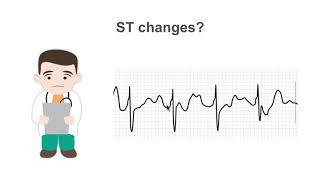
Electrical activities of the heart can be picked up on the skin via electrodes. An ECG machine records these activities and displays them graphically. The graphs show the heart’s OVERALL electrical potential, or voltage, as it changes over time during a cardiac cycle.
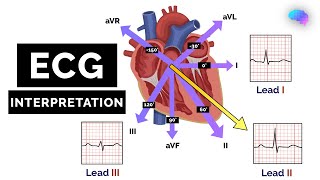
The 12 leads of the ECG represent 12 electrical views of the heart from 12 different angles. The conventional 12lead procedure involves attaching 10 electrodes to the body: one to each limb and six across the chest.
There are 6 limb leads and 6 chest leads.
The 6 limb leads look at the heart in a vertical plane and are obtained from three electrodes attached to the right arm, left arm, and left leg. The electrode on the right leg is an earth electrode.
The measurement of a voltage requires 2 poles: negative and positive. The ECG machine uses the negative pole as zero reference. Thus, the position of the positive pole is the “point of view”, and the line connecting the 2 poles is the “line of sight”.
Leads I, II, and III are BIpolar they measure electrical potential between 2 of the 3 limb electrodes: Lead I represents the voltage between the right arm – negative pole and the left arm – positive pole, and thus looks at the heart from the left. Lead II sees signal movements between the right arm – negative and the left leg –positive forming the INFERIOR LEFT view. Similarly, lead III measures electrical potential between the left arm – negative and the left leg –positive, looking at the heart from an INFERIOR RIGHT angle.
Leads aVR, aVL, and aVF, or “augmented limb leads”, are UNIpolar. They use ONE limb electrode as the positive pole, and take the average of inputs from the OTHER two as the zero reference. Hence, aVR looks at the UPPER RIGHT side of the heart; aVL looks at the UPPER LEFT side of the heart; and aVF looks at the INFERIOR wall of the heart.
The chest leads, or precordial leads, view the heart in a HORIZONTAL plane. These are unipolar leads. The corresponding chest electrodes serve as the positive poles. The reference negative value is the same for all chest leads and is calculated as the average of inputs from the three limb electrodes.
DEpolarization TOWARD a lead produces a POSITIVE deflection; DEpolarization AWAY from a lead gives a NEGATIVE deflection. The REVERSE is true for REpolarization. Thus, leads that look at the heart from different angles may have waves pointing in different directions.