3. P Wave Overview - ECG assessment and ECG interpretation made easy

: https://t.me/bhanuprakashdr
: / drgbhanuprakash
: https://linktr.ee/DrGBhanuprakash
3. P Wave Overview ECG assessment and ECG interpretation made easy
The P wave is the first positive deflection on the ECG and represents atrial depolarisation.
The P wave is the first positive deflection on the ECG
It represents atrial depolarisation
Normal duration: greater than 0.12 s ( greater than 120ms or 3 small squares)
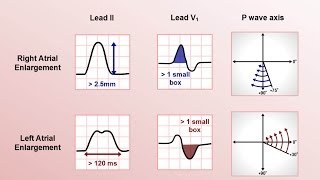
Characteristics of the Normal Sinus P Wave
Morphology
Smooth contour
Monophasic in lead II
Biphasic in V1
Axis
Normal P wave axis is between 0° and +75°
P waves should be upright in leads I and II, inverted in aVR
Duration
Greater than 0.12 s (Greater than 120ms or 3 small squares)
Amplitude
Greater than 2.5 mm (0.25mV) in the limb leads
Greater than 1.5 mm (0.15mV) in the precordial leads
Atrial abnormalities are most easily seen in the inferior leads (II, III and aVF) and lead V1, as the P waves are most prominent in these leads.
The Atrial Waveform – Relationship to the P wave
Atrial depolarisation proceeds sequentially from right to left, with the right atrium activated before the left atrium
The right and left atrial waveforms summate to form the P wave
The first 1/3 of the P wave corresponds to right atrial activation, the final 1/3 corresponds to left atrial activation; the middle 1/3 is a combination of the two
In most leads (e.g. lead II), the right and left atrial waveforms move in the same direction, forming a monophasic P wave
However, in lead V1 the right and left atrial waveforms move in opposite directions. This produces a biphasic P wave with the initial positive deflection corresponding to right atrial activation and the subsequent negative deflection denoting left atrial activation
This separation of right and left atrial electrical forces in lead V1 means that abnormalities affecting each individual atrial waveform can be discerned in this lead. Elsewhere, the overall shape of the P wave is used to infer the atrial abnormality
#pwave #pwaveecg #ecg #ekg #ecgcourse #ecgmadeeasy #ecgonline #ecgreading #ecginterpretation #ekgonline #howtoreadecg #pwaveabnormalities #invertedpwave #enlargedpwave #pwavemorphology #cardiology #normalpwave