3D Kidney Embryology Part 2: Development of Definitive Kidney Nephron u0026 Renal Pelvis

In this 3D visual lecture of Embryology, Dr. Aizaz from MedicoVisual talks about the formation of Kidney.
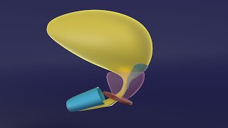
The third, most advanced, last to develop, and caudal most pair kidneys is metanephric kidney. It lies in the pelvic region and forms a mesenchymal condensation called metanephric blastema.
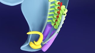
From the caudal end of the mesonephric duct and diverticulum arises that goes towards the metanephric blastema. As it reaches close to the blastema, in its honor, it becomes dilated and gets divided several times.
The last division forms the collecting tubule and each tubule has a small cap of metanephric blastema at its distal end. There are millions of collecting tubules, each wearing its own metanephric cap. The terminal division of the ureteric bud (the diverticulum that arose from the mesonephric duct) induces the development of metanephric blastema, while the metanephric blastema itself causes the ureteric bud to divide and differentiate. This is like a group of two best friends who motivate each other to study hard. Such relationship of inducing each other is called reciprocal induction.
Whole this structure is surrounded by a thin layer of remaining blastema that forms the capsule of this definitive kidney.
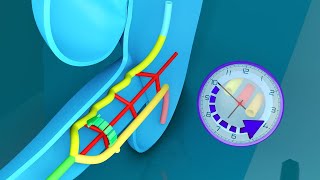
The blastemal cap forms a vesicle by process similar to that of promesonephros. i.e. mesenchyme to epithelium transition and regression of central cells to form cavity. This primitive nephron then assumes an sshape and finally forms a fullfledged nephron. At the medial end, tuft of capillaries invaginate into it to form the bowman’s capsule. The proximal and distal part overgrows and this forms convolutions. These parts of nephron are termed as proximal and distal convoluted tubules respectively. A part of nephron beyond the distal convoluted tubule establishes connection with the collecting duct and is called connecting tubule. From Bowman’s capsule up to the connecting tubule, everything originates from the metanephric blastema while the rest of structures are derived from the ureteric bud (the diverticulum that arises from the mesonephric duct).
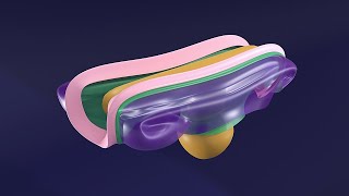
The diverticulum from the mesonephric duct forms the ureter. Its upper dilated part forms the basinlike pelvis of the kidney. Its first division forms the major calyx and next division forms the minor calyces.