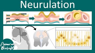
3D Neurulation - Neural Tube formation - Secondary Neurulation - Third Week Embryology

In this visual medical lecture, Dr. Aizaz from MedicoVisual talks about Neurulation or Neural Tube formation. Secondary Neurulation and Primary Neurulation concept is also discussed. Neural tube is the primordium of CNS i.e. Brain and spinal cord. Neurulation starts during the third week of development of the Embryo.
Why Anterior Visceral Endoderm is called "Anterior": • Gastrulation Human Embryology 3rd...
Timestamps:
00:00 Highlights
00:31 Introducing the structures involved
02:17 Neural Plate formation and role of Notochord
04:56 Lengthening of Neural plate and Embryo (as a whole)
05:32 Neural Groove formation / Invagination of Neural Plate
07:26 Fusion of Neural folds and formation of Neuropores
10:03 Function of Neuropores (Anterior and posterior Neurpore)
11:26 Closure of Anterior and Posterior Neuropores
14:05 Crosssection view of Neurulation
16:16 Neuroectoderm vs Surface Ectoderm
16:56 Secondary Neurulation and tail bud
Neurulation Embryology: A Detailed Visual Guide
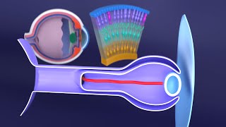
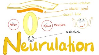
Setting the Stage: The Trilaminar Germ Disc
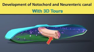
Our story begins with the trilaminar germ disc, the foundation of the developing embryo. For our purposes, we'll focus on two of its layers: the ectoderm, facing the amniotic cavity, and the endoderm, facing the yolk sac cavity. Sandwiched between them lies the crucial notochord, a rodlike structure that plays a pivotal role in neurulation.
The Notochord: The Master Conductor
The notochord isn't just a structural element; it's a signaling powerhouse. It releases chemical factors that act on the overlying ectoderm, inducing its transformation. Think of it as the conductor of an orchestra, directing the ectoderm to thicken and form the neural plate, the first visible sign of our future brain and spinal cord.
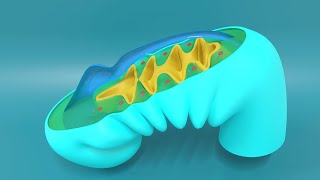
From Plate to Groove: Shaping the Neural Tube
As the embryo grows, so does the neural plate, particularly at its cranial end, destined to become the brain. The plate's edges, marked by future neural crest cells, begin to rise, folding inwards to create the neural groove, a central depression flanked by neural folds. This process resembles folding a flat sheet of paper into a tube.
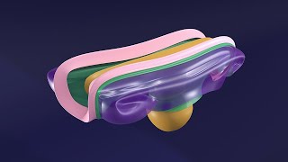
Fusion and Formation: The Neural Tube Takes Shape
The neural folds continue their inward journey, eventually meeting and fusing in the midline. This fusion begins in the future cervical region and progresses both cranially and caudally, like a zipper closing. The once open neural groove is now a closed tube, submerged beneath the surface ectoderm.
Neuropores: Temporary Openings with Vital Roles
Before complete closure, openings at either end of the neural tube, called neuropores, connect the neural tube's interior with the amniotic cavity. These temporary portals are crucial for the developing neural tube, allowing it to receive nutrients and essential factors from the amniotic fluid before its own blood supply is established.
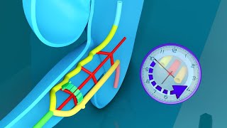
Closure of Neuropores: A Precise Timeline
The cranial neuropore, located at the head end, closes first, around day 2425 of development. The caudal neuropore, at the tail end, follows suit around day 2728. This sequential closure reflects the direction of neural tube fusion, starting cranially and progressing caudally.
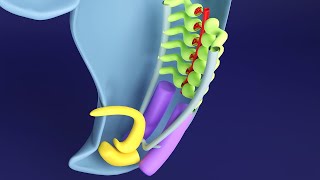
Neural Crest Cells: Ambassadors of the Nervous System
Remember those cells at the neural folds' edges? As the neural tube closes, these neural crest cells detach and migrate throughout the body. These remarkable cells differentiate into a diverse array of structures, including components of the peripheral nervous system, pigment cells in the skin, and even parts of the face and skull.
Primary vs. Secondary Neurulation: Two Paths to One Goal
What we've discussed so far is primary neurulation, the main mechanism for neural tube formation. However, a secondary process, aptly named secondary neurulation, occurs in the tail bud, the region beyond the notochord's reach.
Here, mesenchymal cells aggregate to form a solid cord called the medullary cord. Cells in the cord's center undergo programmed cell death, creating a hollow cavity. This newly formed secondary neural tube then fuses with the primary neural tube, ensuring a continuous structure from head to tail.
Secondary Neurulation in Humans: A Limited Role
While crucial in some species, secondary neurulation's contribution in humans is debated. Some evidence suggests it might be involved in forming the lowermost part of the spinal cord, the sacrococcygeal region.
Conclusion
Neurulation is a beautifully orchestrated process, transforming a flat sheet of cells into the foundation of our central nervous system. Understanding this intricate dance of signaling, folding, and fusion is essential for comprehending both normal development and the potential consequences of developmental errors. Thank you for joining me on this journey through the fascinating world of neurulation.
Website: https://www.medicovisual.com
Email: [email protected]


![Gastrulation Human Embryology 3rd Week [Animated] MedicoVisual](https://i.ytimg.com/vi/0n_Ev8DbQgY/mqdefault.jpg)