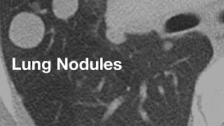
Acute Epiploic Appendagitis | Radiology Board Review Case

CT appearance of Acute Epipolic Appendagitis and its differentials.
Subscribe for more radiology content like this: https://bit.ly/radiogyanYT
Full post and further reading: https://radiogyan.com/videos/acuteep...
Acute epiploic appendagitis presents as acute lower quadrant pain. Clinical features are similar to those of acute diverticulitis or, less commonly, acute appendicitis.
Differentials include acute omental infarction, mesenteric panniculitis, fatcontaining tumor, and primary and secondary acute inflammatory processes in the large bowel.
The CT features of acute epiploic appendagitis is that of an oval lesion 1.5–3.5 cm in diameter, with fat attenuation , surrounding inflammatory changes, abutting the anterior sigmoid colon wall.
This is a self limiting condition.
Join our #radiology discussion groups to participate in the discussion live:
Telegram: https://t.me/radiogyan
WhatsApp: https://radiogyan.com/recommends/radi...
Website https://radiogyan.com
Recommended Radiology Books: https://radiogyan.com/radiologyresou...
Sign up to my monthly email newsletter https://radiogyan.com/recommends/radi...
Instagram / radiogyan
Twitter / radiogyan
Facebook / radiogyan
Donate: https://paypal.me/radiogyan
♂ WHO AM I:
I'm Amar Udare, a radiology fellow training at McMaster University Canada. I have a passion for teaching and I believe in #FOAMrad Free Open Access Medicine Radiology.
WHAT CONTENT CAN YOU EXPECT?
I share radiology cases and resources which will help you prepare for your radiology board exams (including MD / DMRD / DNB radiodiagnosis, the #FRCR exam, OSCE based radiology exams and other radiology exam patterns).
GET IN TOUCH:
If you’d like to talk, I’d love to hear from you. Messaging @radiogyan on Twitter directly will be the quickest way to get a response, but if your question is very long, feel free to email me at [email protected]. I try my best to reply to everyone.
⏱Time Stamps:
0:00 Introduction
0:40 PACS based case images.
1:00 Description of imaging findings.
3:10 Pathophysiology of acute epiploic appendagitis.
10:30 Discussion of imaging findings and differentials for acute epiploic appendagitis .
DISCLAIMER: Some of the links included in this description might be affiliate links. If you purchase a product or service with the links that I provide I may receive a small commission. As an Amazon Associate, I earn from qualifying purchases. There is no additional charge to you! Thank you for supporting my channel so I can continue to provide you with free content each week!
All Rights Reserved.
No part of the content of this video or any video published under RadioGyan, and other encompassed entities, shall be stored, copied, recreated, republished, or transported. Prior express written permission by RadioGyan or Amar Udare is required for any use of this video not permitted the Copyright Act.
The advice, guidance, and principles outlined in all content produced by RadioGyan, and other encompassed entities are not guaranteed to be appropriate for your unique situation. We recommend that as a consumer, you exercise your due diligence and research any and all strategies recommended to you before adopting them. RadioGyan and encompassed entities are not responsible for any damages that result from an effort to implement the information provided in this or any other video, article, social media post, and related publications. Your use and viewing of any materials and videos published by RadioGyan, and other encompassed entities confirms your acknowledgement and agreement that Indian law will apply to any and all disputes related to the aforementioned entities.
The information in the videos is for educational purpose only and is intended for medical professionals. It should not be used for selfdiagnosis or selftreatment. It is not intended as, nor should it be, a substitute for independent professional medical care. We recommend that you consult your own physician regarding any diagnosis, imaging interpretation or course of treatment. Medical practitioners must make their own independent assessment before suggesting a diagnosis or recommending or instituting a course of treatment. The Platforms should not in any way be seen as a replacement for consultation with colleagues or other sources, or as a substitute for conventional training and study.
If you are not medically qualified and are registering as a layperson, you further acknowledge that the content on the Platforms is provided for educational purposes only, and is provided for use by medical professionals. You agree to use the information solely for your own private educational purposes and further agree not to rely on the information in any way.


![Radiology Board Core Exam Review Cases [Quiz and Discussion] | Set 10 | Sep 2020](https://i.ytimg.com/vi/uuCRFjAy86s/mqdefault.jpg)