Anatomy Lateral Femoral Cutaneous Nerve Of Thigh - Everything You Need To Know - Dr. Nabil Ebraheim

Dr. Ebraheim’s educational animated video describes the Anatomy of the lateral Femoral Cutaneous Nerve of the Thigh in a very easy and simple animation.
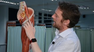
The lateral femoral cutaneous nerve of the thigh arises from the dorsal branches of L2 and L3 and the course of the nerve can be variable.
With its origin coming from the nerve roots of L2 and L3, the lateral femoral cutaneous nerve innervates the skin on the lateral part of the thigh.
The LFCM can become injured during:
•Harvesting anterior iliac crest bone graft.
•Ilioinguinal approach for acetabulum fixation.
•Application of external fixator if the pelvis.
•Total hip replacement by anterior approach or smith Peterson approach.
The LFCN usually passes under the inguinal ligament approximately 2 cm medial to the ASIS. Once outside the pelvis, the nerve splits and pierces the fascia, running over the lateral aspect of the thigh in the subcutaneous region and lies superficial to the Sartorius' muscle. These variations can cause entrapment of the nerve near the iliac crest called meralgia paresthetica.
Recognizing the variability and the relationship of this nerve and its branches in relationship to specific anatomic landmarks is important to avoid injury to the nerve. It is important in order to establish a “safe zone” during surgical approaches to the acetabulum or proximal femur.
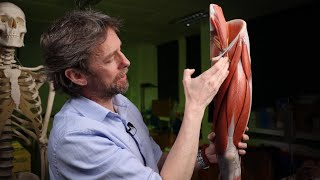
Determine the distance of the LFCN and its branches medial from the anterior superior iliac spine. The angle that the LFCN and its branches make with the inguinal ligament (x).
The LFCN crosses the lateral border of the Sartorius' muscle at a mean of 54 mm inferior and 12 mm medial (c) to the ASIS.
The LFCN divides into anterior and posterior divisions on the surface of the Sartorius approximately 5 cm below the ASIS in most patients. Approximately 27.6% of the nerve studied had divided before crossing the inguinal ligament.
The distance that the branches of the LFCN crossed the inguinal ligament ranged from 6 mm to 7.3 cm medial to the ASIS along the inguinal ligament.
The distance that the branches of the LFCN crossed the Sartorius inferior (B) to the ASIS ranged from 2.2 cm inferior to 11.3 cm along the lateral border of the sartorius.
The distance that the nerve branches crossed the lateral border of the sartorius (C) is shown in reference to a line drawn vertically from the ASIS.the nerve branches cross this area at an average of 128 mm medial to this line.
The distance along the inguinal ligament (a) that the LFCN and its branches crossed ranged from 6 mm 7.3 mm medially along the inguinal ligament.
The distance extends inferiorly (b) along the lateral border of the Sartorius, which the nerve and its branches cross, from 2.2 cm inferior to the ASIS to 11.3 cm distal. Line (c) is the distance that the branch of the LFCN with the most medial distance from the ASIS extends to the line drawn vertically from the ASIS. Line (d) is used to complete the perimeter of the area at risk of injury during surgery
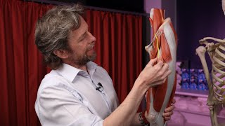
The usual traumatic site for the LFCN is at the inguinal ligament.
Become a friend on facebook:
/ drebraheim
Follow me on twitter:
https://twitter.com/#!/DrEbraheim_UTMC