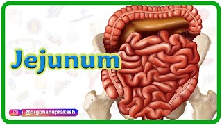
Anatomy of Cecum Animation : Relations Blood supply Venous drainage Nerve supply and development

: / drgbhanuprakash
: https://t.me/bhanuprakashdr
: https://linktr.ee/DrGBhanuprakash
Anatomy of Cecum Animation : Relations, Blood supply, Venous drainage, Nerve supply and development
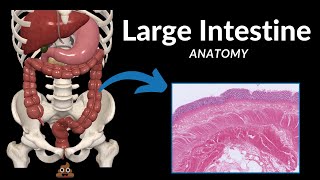
The cecum is a pouchlike structure that forms the beginning of the large intestine. It is located in the lower right quadrant of the abdomen, in the region known as the right iliac fossa. The cecum plays a role in the absorption of fluids and salts and serves as a site for the fermentation of indigestible material by bacteria.
Here's an animated description of the anatomy of the cecum, including its relations, blood supply, venous drainage, nerve supply, and development:
Relations:
The cecum is situated in the lower right quadrant of the abdomen, just below the ileocecal junction.
It lies anterior to the iliacus muscle and the psoas major muscle.
The terminal part of the ileum (the last portion of the small intestine) attaches to the medial side of the cecum at the ileocecal valve.
The cecum is usually located in close proximity to the appendix, which is a fingerlike projection from its posteromedial aspect.
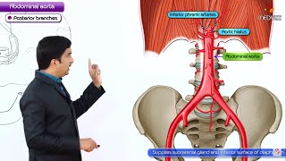
Blood Supply:
The arterial blood supply to the cecum comes from the ileocolic artery, a branch of the superior mesenteric artery.
The ileocolic artery runs posterior to the terminal ileum, and its branches supply blood to the cecum and appendix.
In some individuals, there may be additional blood supply from the anterior cecal artery, which arises from the appendicular artery or the posterior cecal artery, arising directly from the ileocolic artery.
Venous Drainage:
Venous blood from the cecum drains into the superior mesenteric vein, which ultimately merges with the splenic vein to form the hepatic portal vein.
The venous drainage pattern generally parallels the arterial supply, with the ileocolic vein receiving blood from the cecum and appendix.
Nerve Supply:
Innervation of the cecum is primarily provided by the superior mesenteric plexus, which consists of sympathetic and parasympathetic fibers.
Sympathetic fibers originate from the superior mesenteric ganglion, while parasympathetic fibers come from the vagus nerve.
Sensory innervation of the cecum is mediated by visceral afferent fibers.
Development:
During embryonic development, the cecum arises as an outpouching of the primitive hindgut.
It undergoes rotation during fetal development, assuming its final position in the right lower quadrant of the abdomen.
The appendix, which is derived from the cecum, develops as a tubular structure extending from the posteromedial aspect of the cecum.
#anatomyvideos #anatomy #cecumanatomy #abdomenandpelvis #abdomenanatomy #abdomen #fmge #fmgevideos #rapidrevisionfmge #fmgejan2023 #mbbslectures #nationalexitexam #nationalexittest #neetpg #usmlepreparation #usmlestep1 #fmge #usmle #drgbhanuprakash #medicalstudents #medicalstudent #medicalcollege #neetpg2023 #usmleprep #usmlevideos #usmlestep1videos #medicalstudents #neetpgvideos #cecum