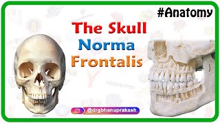
Anatomy of Frontal Bone - Animated Osteology MBBS 1st year

: / drgbhanuprakash
: https://t.me/bhanuprakashdr
: https://linktr.ee/DrGBhanuprakash
Frontal bone
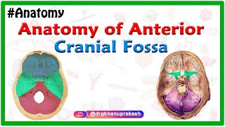
The frontal bone is a skull bone that contributes to the cranial vault. It contributes to form part of the anterior cranial fossa.
Gross anatomy
The frontal bone has two portions:
vertical portion (squama): has external/internal surfaces
horizontal portion (orbital): has superior/inferior surfaces
External surface of the vertical portion features:
frontal/metopic suture
frontal eminence (tuber frontale)
superciliary arches which join to form the glabella
supraorbital margin
supraorbital notch/foramen containing the supraorbital nerve and vessels
supratrochlear notch/foramen containing the supratrochlear nerve and vessels
zygomatic process laterally
upper and lower temporal lines run backwards and are the sites of attachment for the temporalis fascia and temporalis muscle, respectively
nasal part features the nasal notch, nasion (middle of frontonasal suture), and nasal process (sharp spine, forms part of nasal septum)
coronal suture
bregma
pterion
Internal surface of the vertical portion features:
sagittal sulcus: vertical groove for the superior sagittal sinus
frontal crest: ridge, formed from edges of sulcus that gives attachment to the falx cerebri
foramen caecum: small notch, converted into foramen, emissary vein from nose
groove for anterior meningeal artery (branch of anterior ethmoidal artery)
The horizontal portion is composed of two thin, orbital plates separated by the ethmoidal notch.
Inferior surface of the horizontal portion is smooth and concave. It features:
lacrimal fossa: lateral shallow depression for lacrimal gland
fovea trochlearis / trochlear spine near nasal part, for attachment of cartilaginous pulley for superior oblique muscle
Superior surface of the horizontal portion is convex and contains depressions for cerebral convolutions. It features:
openings for the frontal sinuses on either side of the nasal process; each frontal sinus communicates with the ipsilateral middle nasal meatus via a frontonasal duct
two grooves converted into anterior and posterior ethmoidal canals when articulating with the ethmoid bone
#frontalbone #frontalboneanatomy #frontalboneanimation #frontalboneosteology #animatedanatomy #frontalboneusmle #frontalbonelecture #frontalbonevideo #drgbhanuprakash #usmle #usmlestep1 #animatedanatomylectures #animatedanatomyvideos #drgbhanuprakashanatomyvideos


![Skull Bone & Suture Mnemonic/Trick [Cranial Bone Anatomy Animation]](https://i.ytimg.com/vi/P21JpzKtlxE/mqdefault.jpg)


![Facial Bones of the Skull Mnemonic [Anatomy Animation]](https://i.ytimg.com/vi/d2D-OMUD2q0/mqdefault.jpg)