Anatomy of Hip Bone / innominate bone / Pelvis ( Osteology ): Ilium Ischium Pubis: Animation

: / drgbhanuprakash
: https://t.me/bhanuprakashdr
: https://linktr.ee/DrGBhanuprakash
Anatomy of Hip Bone / innominate bone ( Osteology ) : Ilium , Ischium , Pubis
The pelvis is a bony structure that can be found in both male and female skeletons. The exception to this compound structure, when compared to all other bones, is that it has differences that are classified by sex, both for functional and general developmental reasons. The rest of the human skeleton differs only in size, which is genetically determined and is usually slightly larger in males than in females.
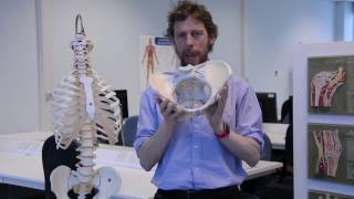
The pelvis is formed by four bones which include a pair of hip bones otherwise known as innominate bones, the sacrum, which comes with the five lower sacral bones that are fused together and the coccyx which has four fused and a single individual terminal vertebra. The pelvic girdle consists of the hip bones and the sacrum and its function is to transmit the weight from the upper body to the lower limbs while allowing the body to stay balanced. Meanwhile, the ilium, the ischium, and the pubis fuse together at puberty to form the innominate bones and are joined by the cartilage found in the acetabulum.
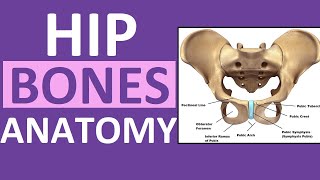
The left and right hip bones (innominate bones, pelvic bones) are two irregularly shaped bones that form part of the pelvic girdle – the bony structure that attaches the axial skeleton to the lower limbs.
The hip bones have three main articulations:
Sacroiliac joint – articulation with the sacrum.
Pubic symphysis – articulation between the left and right hip bones.
Hip joint – articulation with the head of the femur.
In this article, we shall look at the anatomy of the hip bones – their composition, bony landmarks, and clinical relevance.
Composition of the Hip Bone
The hip bone is comprised of the three parts; the ilium, pubis, and ischium. Prior to puberty, the triradiate cartilage separates these parts – and fusion only begins at the age of 1517.
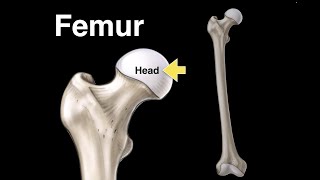
Together, the ilium, pubis, and ischium form a cupshaped socket known as the acetabulum (literal meaning in Latin is ‘vinegar cup‘). The head of the femur articulates with the acetabulum to form the hip joint.
The Ilium
The ilium is the widest and largest of the three parts of the hip bone and is located superiorly. The body of the ilium forms the superior part of the acetabulum (acetabular roof). Immediately above the acetabulum, the ilium expands to form the wing (or ala).
The wing of the ilium has two surfaces:
Inner surface – has a concave shape, which produces the iliac fossa (site of origin of the iliacus muscle).
External surface (gluteal surface) – has a convex shape and provides attachments to the gluteal muscles.
The superior margin of the wing is thickened, forming the iliac crest. It extends from the anterior superior iliac spine (ASIS) to the posterior superior iliac spine (PSIS).
On the posterior aspect of the ilium, there is an indentation known as the greater sciatic notch.
The Pubis
The pubis is the most anterior portion of the hip bone. It consists of a body, superior ramus and inferior ramus (ramus = branch).
Pubic body – located medially, it articulates with the opposite pubic body at the pubic symphysis. Its superior aspect is marked by a rounded thickening (the pubic crest), which extends laterally as the pubic tubercle.
Superior pubic ramus – extends laterally from the body to form part of the acetabulum.
Inferior pubic ramus – projects towards the ischium.
Together, the superior and inferior rami enclose part of the obturator foramen – through which the obturator nerve, artery, and vein pass through to reach the lower limb.
The Ischium
The ischium forms the posteroinferior part of the hip bone. Much like the pubis, it is composed of a body, an inferior ramus, and superior ramus.
The inferior ischial ramus combines with the inferior pubic ramus forming the ischiopubic ramus, which encloses part of the obturator foramen. The posteroinferior aspect of the ischium forms the ischial tuberosities and when sitting, it is these tuberosities on which our body weight falls.
Near the junction of the superior ramus and body is a posteromedial projection of bone; the ischial spine.
Two important ligaments attach to the ischium:
Sacrospinous ligament – runs from the ischial spine to the sacrum, thus creating the greater sciatic foramen through which lower limb neuro vasculature (including the sciatic nerve) transcends.
Sacrotuberous ligament – runs from the sacrum to the ischial tuberosity, forming the lesser sciatic foramen.
#hipboneanatomy #pelvicbone #innominatebone #animatedanatomy #drgbhanuprakash #anatomyanimations #anatomy #hipbone #AnatomyofHipBone #