Anatomy of the Heart - Layers Conducting System u0026 Topography

Content:
0:00 Introduction
0:29 Layers of the Heart
1:12 Endocardium
2:04 Myocardium
5:18 Epicardium
5:59 Pericardium
7:21 Cardiac Conduction System
8:34 Topography of the Heart
Channel membership: / @taimtalksmed
Follow my IG: / taimtalksmed
Donation link: https://www.buymeacoffee.com/taimtalk...
Layers of the Heart:
Serous Pericardium
Fibrous Pericardium
Endocardium
Myocardium
Epicardium
Endocardium
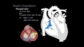
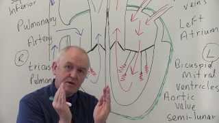
Forms the Cusps of the:
○ Bicuspid Valve
○ Tricuspid Valve
○ Aortic Valve
○ Pulmonary Valve
○ Valve of the inferior Vena Cava (Vulva Vena Cava Inferioris)
○ Valve for the Coronary Sinus (Vuvla Sinus Coronarii)
Myocardium
Form Fibrous Rings
Myocardium of the Atria
○ Superficial Circular Muscle Fibers
○ Deep Longitudinal Muscle Fibers
○ Form Pectinate Muscle (Musculi Pectinati)
Myocardium of the Ventricles
○ Deep Layer: Longitudinal Muscle Fibers
○ Middle Layer: Circular Muscle Fibers
○ Superficial Layer: Longitudinal Muscle Fibers
○ Deep and Superficial muscle fibers are connected, they form the Vortex of the Heart (Vortex Cordis)
○ Left Ventricle thicker than Right Ventricle
○ Form Trabeculae Carneae
○ Papillary Muscles (Musculi Papillares)
Epicardium
It's the Visceral Lamina of the Serous Pericardium
Pericardium
Serous Pericardium (Pericardium Serosum)
○ Visceral Lamina (Epicardium)
○ Parietal Lamina
Fibrous Pericardium (Pericardium Fibrosum)
Pericardial Cavity (Cavitas Pericardiaca)
○ Transverse Pericardial Sinus (Sinus Transversus Pericardii)
○ Oblique Pericardial Sinus (Sinus Obliquus Pericardii)
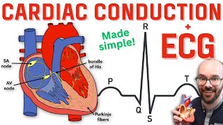
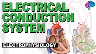
Conducting System of the Heart:
Sinoatrial Node (SA Node)
Atrioventricular Node (AV Node)
Bundle of His
Left Bundle Branch
Right Bundle Branch
Purkinje Fibers
Topography of the heart:
Holotopy of the heart:
○ Heart lies in the Mediasternum Medium
Skeletopy of the heart:
Superior Border: Third rib horizontally
Right Border: Parallel to sternal margin. Linea Parasternalis
Lower Border: Obliquely: From Cartilage of 5th rib to 5th intercostal space.
Left Border: 5th intercostal space to the level of the 4th rib.
Openings op the heart:
Atrioventricular Openings: 3rd rib to the 6th sternal junction.
Aortic and Pulmonary openings: 3rd sternal junction to the 4th sternal junction
Topography of the valves of the heart:
Aortic and Pulmonary Valve: 2nd intercostal Space
Tricuspid and Bicuspid Valve: 5th intercostal space.
Syntopia of the heart:
Anteriorly: Behind Sternum
Posteriorly: Oesophagus
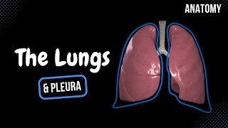
Laterally: Pleura
Inferiorly: Diaphragm
Superiorly: Great Blood vessels
Sources used in this video:
Memorix Anatomy 2nd Edition by Hudák Radovan (Author), Kachlík David (Author), Volný Ondřej (Author)
Biorender
University notes and lectures