Anatomy Of The Peroneal Muscles In The Lower Leg - Everything You Need To Know - Dr. Nabil Ebraheim

Dr. Ebraheim’s educational animated video describes the anatomy of the Peroneal muscles in the lower leg, with simple images; this video also provides you with all you need to know about this muscle, its innervation, action, and function.
The peroneal muscles are a group of three muscles:
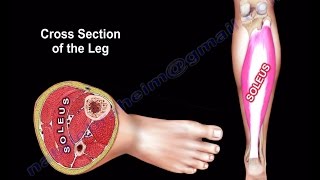
The peroneus longus, peroneus brevis, and the peroneus tertius. Others may exist. The peroneus longus and brevis muscles lie within the lateral compartment and the peroneus tertius muscle in located within the anterior compartment of the leg. The peroneus brevis muscle arises from the lower 2/3 of the lateral surface of the fibula. The peroneus tertius muscle arises from the lower ¼1/3 of the anterior portion of the medial surface of the fibula. The peroneus longus is the longest and most superficial muscle of the lateral compartment. The tendon of the peroneus longus muscle begins at a higher level than the tendon of the peroneus brevis and can easily be recognized on ultrasound. The peroneus longus is lateral and posterior to the peroneus brevis muscle. Near the ankle and on the ultrasound image, the peroneus longus appears as a tendon while the peroneus brevis may appear as a muscle.
The peroneus longus muscle is inserted into two bones:
•The base of the first metatarsal
•Adjoining portion of the medial cuniform bone
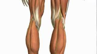
Before the peroneus longus insertion, the tendon makes three turns. The first turn is at the tip of the lateral malleolus. The second turn occurs below the trochlear process of the calcaneous. Finally, it turns at the groove of the cuboid crossing the plantar surface of the foot obliquely. The peroneus brevis muscle is inserted into the tuberosity of the base of the 5th metatarsal bone. The peroneus brevis muscle can be used as a flap to reconstruct a small defect of the distal third of the lower leg. The peroneus tertius muscle is inserted into dorsal surface of the base of the 5th metatarsal bone. The location of the anterolateral ankle arthroscopy portal should lie just lateral to the peroneus tertius tendon. Staying lateral to the peroneus tertius helps avoid injury to the dorsal lateral branch of the peroneal nerve. The peroneal muscles are situated on the outer side of the lower leg and their tendons attach to the foot. Nera the ankle, the peroneus brevis is closer to the fibula.
There are two peroneal retinacula which hold the two peroneal tendons: Superior peroneal retinaculum (important) and inferior peroneal retinaculum. Rupture of the superior retinaculum may cause peroneal subluxation and the subluxation may be acute, chronic, or recurrent. Acute rupture of the superior peroneal retinaculum allows for subluxation of the peroneal tendons and may cause disability of the ankle. Retromalleolar pain on active eversion is a specific and highly suggestive finding for dislocation of the peroneal tendon. Injury to the peroneal tendons is a frequently overlooked cause of persistent lateral ankle pain after trauma. The most reliable sign is persistent swelling along the posterolateral edge of the fibula.
A pathognomonic sign for peroneal tendon subluxation is an avulsion of a piece of bone from the fibula. The “fleck sign” is an indication for peroneal tendon subluxation. An xray may show an avulsion fracture of the fibula called a rim fracture. The piece of bone from the avulsion fracture is long and thin. Tear of the superior retinaculum may be misdiagnosed because of the associated pain, swelling, and ecchymosis that may hinder early diagnosis.
Follow me on twitter:
https://twitter.com/#!/DrEbraheim_UTMC
Background music provided as a free download from YouTube Audio Library.
Song Title: Every Step