Anatomy Of The Popliteal Fossa - Everything You Need To Know - Dr. Nabil Ebraheim

Dr. Ebraheim’s educational animated video describes the anatomy associated with the popliteal fossa posterior knee.
Follow me on twitter:
https://twitter.com/#!/DrEbraheim_UTMC
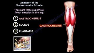
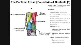
The area of depression located at the back of the knee joint is called the popliteal fossa. The popliteal fossa is a closelypacked space. It is bounded by the biceps femoris laterally, as well as the semitendinosus and semimembranosus medially. The lower part of the space is formed by the two heads of the gastrocnemius muscle.
Four common conditions involving the popliteal fossa include:
1. Baker’s Cyst
2. Popliteal Artery Entrapment Syndrome
3. Posterior Knee Dislocation
4. Posterior Cruciate Ligament (PCL) Injury
A Baker’s cyst is a benign swelling found behind the knee that lies between the semimembranosus and the medial gastrocnemius muscles. A Baker’s cyst is also known as a popliteal cyst which lies posterior to the medial femoral condyle. The cyst is connected to the knee joint through a valvular opening. Knee effusion from intraarticular pathology allows the fluid to go through the valve to the cyst in one direction.
Popliteal artery entrapment syndrome is a rare condition involving extrinsic compression of the popliteal artery behind the knee due to the anomalous relationship of the muscle and artery in the popliteal fossa. It may also be caused by fibrous tissue constricting the artery. This condition usually affects younger athletes who present with calf claudication. The blood flow will be decreased. The patient will complain of swelling, foot numbness and paresthesia, tingling of the toes, and cramping of the muscles. Plantar flexion of the ankle and hyperextension of the knee will decrease the pulses. An arteriogram is probably the best study showing the compression and condition of the artery. Treatment consists of observation and activity modification. Surgery may be necessary to release the muscle and relieve the pressure on the artery.
Posterior knee dislocation occurs as a result of violent trauma. The most common mechanism of injury includes exaggerated hyperextension of the knee and dashboard injuries. Posterior knee dislocation may be associated with a high incidence of popliteal artery injury.
Posterior translation of the tibia will occur with rupture of the posterior cruciate ligament. A common cause of this injury is a bent knee hitting a dashboard during a car accident; however, it occurs more frequently in sports from forced hyperflexion of the knee.
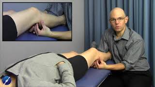
The quadriceps active test is conducted to test the quadriceps muscle. The examiner stabilizes the leg of the patient and then the patient is asked to actively contract the quadriceps muscle. The tibia is seen being actively reduced from the posterior subluxed position. The Lachman’s Test is another test used to examine the knee. When performing the Lachman’s test, the knee is bent at 2030°. The examiner provides posterior force to the tibia while applying anterior pressure to the femur in order to access the posterior translation of the tibia. The posterior drawer test is carried out while the patient is in a supine position and the knee is flexed to 90°. The amount of translation of the tibia relative to the femur is observed. The dial test is performed while the patient is in the supine or prone position and both knee are in 90° (it shows the PCL injury) and 30° of flexion (will show the posterolateral corner injury). More than 10 degrees of external rotation indicates significant injury.