Anatomy Of The Quadriceps - Everything You Need To Know - Dr. Nabil Ebraheim

Dr. Ebraheim’s educational animated video describes the anatomy of the quadriceps muscle group.
The quadriceps femoris is a large muscle group which includes the four muscles that cover the frond and sides of the femur. The quadriceps is divided into four heads:
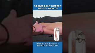
1Vastus lateralis
2Vastus intermedius
3Vastus medialis
4Rectus femoris
The rectus femoris is an important muscle with two heads originating from the pelvis
1Straight head arises from the anterior inferior iliac spine
2Reflected head arises from the groove above the rim of the acetabulum.
The rectus femoris muscle is the only one of the quadriceps muscles that cross the hip joint. The other muscles of the quadriceps do not cross the hip joint.
An avulsion fracture may occur from a strong contraction of the rectus femoris muscle, pulling the tendon and a small piece of bone away from the attachment point. The rectus femoris muscle continues down the thigh and is then inserted into the top of the patella.
The quadriceps muscles are supplied by the femoral nerve. The femoral nerve is the largest branch of the lumbar plexus. The femoral nerve arises from the 2nd, 3rd, & 4th lumbar nerves (L2L4).
Function: extension of the knee. Injury to the femoral nerve will cause atrophy of the quadriceps muscles and loss of function. The patient will be unable to straighten the knee. The quadriceps is inserted into the patella. From the patella, the quadriceps tendon becomes the patellar tendon which then attaches to the tibia. Rupture of the quadriceps tendon is usually associated with intense pain and occurs just above the superior pole of the patella.
Quadriceps tendon rupture
•Occurs more in males and older age groups
•Occurs more in patients with metabolic diseases such as gout, diabetes, renal failure & hyperthyroidism.
Examination:
•swelling at or above the knee.
•Palpable defect above the kneecap.
An xray will show the kneecap is moved distally due to the pull of the patellar tendon
MRI showing the rupture of the quadriceps tendon.
Contracture of the knee
•Unable to bend the knee due to tight quadriceps muscles and tendons form adhesion.
•Treatment of contracture includes physical therapy, manipulation of the knee or possible surgery.
Synovial fluid is a normal fluid inside the knee. A minimal amount of fluid in the knee is normal. The normal amount of fluid within the knee is about 2 ml and when the fluid increases for any reason such as infection or trauma, the quadriceps will not function properly. 20 cc or fluid or effusion will cause the quadriceps muscle inhibition (dysfunction)
Become a friend on facebook:
/ drebraheim
Follow me on twitter:
https://twitter.com/#!/DrEbraheim_UTMC