CARTILAGE - Histology Types Functions

Cartilage is an avascular, smooth and elastic tissue that consists of chondrocytes and an extensive extracellular matrix.
Chondrocytes are specialized cells that produCartilage is an avascular, smooth and elastic tissue that consists of chondrocytes and an extensive extracellular matrix.
Chondrocytes are specialized cells that produce and maintain the extracellular matrix. Extracellular matrix is the extracellular part of multicellular structure that typically provides structural and biochemical support to the surrounding cells.
So what are the functions of cartilages ?
It can act as a smooth surface for bone articulation within joints, play a role in bone formation, and also provide structural support, such as the cartilage rings in the trachea.
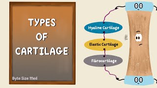
Now, there are three types of cartilage that differ in appearance and mechanical properties. Hyaline cartilage, elastic cartilage, and fibrocartilage.
Let’s first focus on hyaline cartilage, which is the most common type of cartilage.
Hyaline cartilage forms a thick layer over bone ends in joints. And it is also found in the ribs, the septum of the nose which separates the nostrils, and the trachea.
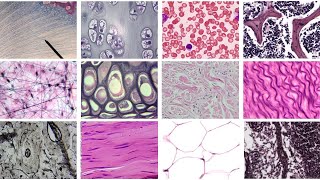
Let's take a closer look at the structure of hyaline cartilage.
This image is a longitudinal section of the trachea, which is an example of hyaline cartilage providing structural support.
Hyaline cartilage is covered externally by a fibrous membrane known as the perichondrium.
Perichondrium consists of an outer fibrous layer and an inner chondrogenic layer.
In the inner chondrogenic layer, we can see some spindleshaped cells. These spindleshaped cells have the capability to differentiate into chondroblasts. So what are chondroblasts ?
Chondroblasts are the precursors of the chondrocytes which are the mature cartilage cells. And also chondroblasts contribute to the formation of the extracellular matrix.
As we mentioned before chondrocytes are mature cartilage cells so where are they found in hyaline cartilage?
There are spaces called lacunae throughout the extracellular matrix.
And chondrocytes are located within these lacunae. And chondrocytes in hyaline cartilage are often arranged in clusters of 2 to 4 cells.
Extracellular matrix of hyaline cartilage has a glassy appearance and it is composed of mostly type II collagen and proteoglycan.
And hyaline cartilage that covers the articular surfaces of movable joints is termed articular cartilage. In general, the structure of articular cartilage is similar to that of hyaline cartilage. However, the free, or articular, surface has no perichondrium.
Now lets talk about the elastic cartilage.
Elastic cartilage is found in the epiglottis, larynx, and the pinna of the external ear.This image is from the pinna of the external ear.
We can see some general features of elastic cartilage that are similar to hyaline cartilage, such as the cartilage matrix, which stains blue; the chondrocytes within lacunae; and also the perichondrium at the top, which is stained purple and dark blue.
However, unlike hyaline cartilage, elastic cartilage has abundant network of elastic fibers which gives this type of cartilage its flexibility and elastic characteristics.
And finally, fibrocartilage. Fibrocartilage is a combination of dense regular connective tissue and hyaline cartilage.
There is also no surrounding perichondrium as in hyaline and elastic cartilage.
Fibrocartilage is found in structures such as the meniscus and intervertebral discs.