Clavicle Bone Anatomy: Bony Landmarks and Articulations Functions Attachments Clinical aspects

: / drgbhanuprakash
: https://t.me/bhanuprakashdr
: https://linktr.ee/DrGBhanuprakash
Clavicle Bone Anatomy : Bony Landmarks and Articulations , Functions, Attachments, Clinical aspects
The clavicle (collarbone) extends between the manubrium of the sternum and the acromion of the scapula.
It is classed as a long bone and can be palpated along its length. In thin individuals, it is visible under the skin. The clavicle has three main functions:
Attaches the upper limb to the trunk as part of the ‘shoulder girdle’.
Protects the underlying neurovascular structures supplying the upper limb.
Transmits force from the upper limb to the axial skeleton.
In this article, we shall look at the anatomy of the clavicle – its bony landmarks and clinical correlations.
Bony Landmarks and Articulations
The clavicle is a slender bone with an ‘S’ shape. Facing forward, the medial aspect is convex, and the lateral aspect concave. It can be divided into a sternal end, a shaft and an acromial end.
Sternal (medial) End
The sternal end contains a large facet – for articulation with the manubrium of the sternum at the sternoclavicular joint.
The inferior surface of the sternal end is marked by a rough oval depression for the costoclavicular ligament (a ligament of the SC joint).
Shaft
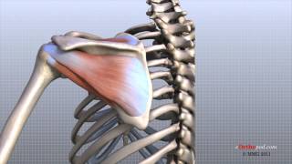
The shaft of the clavicle acts a point of origin and attachment for several muscles – deltoid, trapezius, subclavius, pectoralis major, sternocleidomastoid and sternohyoid
Acromial (lateral) End
The acromial end houses a small facet for articulation with the acromion of the scapula at the acromioclavicular joint. It also serves as an attachment point for two ligaments:
Conoid tubercle – attachment point of the conoid ligament, the medial part of the coracoclavicular ligament.
Trapezoid line – attachment point of the trapezoid ligament, the lateral part of the coracoclavicular ligament.
The coracoclavicular ligament is a very strong structure, effectively suspending the weight of the upper limb from the clavicle.
Clinical Relevance: Fracture of the Clavicle
The clavicle acts to transmit forces from the upper limb to the axial skeleton. Given its relative size, this leaves it particularly susceptible to fracture. The most common mechanism of injury is a fall onto the shoulder or onto an outstretched hand.
With the clavicle arbitrarily divided into thirds:
15% of fractures occur in the lateral third
80% occur in the middle third
5% occur in the medial third.
After a fracture, the lateral end of the clavicle is displaced inferiorly by the weight of the arm and displaced medially by the pectoralis major. The medial end is pulled superiorly by the sternocleidomastoid muscle.
Management of a clavicular fracture can be conservative (e.g. sling immobilization) or operative (e.g. open reduction and internal fixation). The supraclavicular nerves lie in close proximity to the clavicle and are occasionally sacrificed during a surgical repair – resulting in a numb patch over the upper chest and shoulder.
#clavicle #claviclebone #clavicleanatomy #clavicularfracture #clavicleosteology #clavicleattachments #claviclefracture #osteologyofclavicle #anatomyofclavicle #humananatomy #mbbsanatomy #mbbs1styear #usmle #bpt #usmlestep1 #usmlestep2ck #plab #fmge #neetpg #nationalexittext #nationalexitexam #medicalschool #medicalstudents #neetpg #usmlestep1 #clavicleanatomy


![Anatomical Position and Directional Terms [Anatomy MADE EASY]](https://i.ytimg.com/vi/t6-ueqFK1IE/mqdefault.jpg)