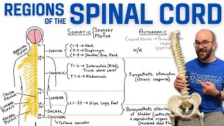
CN 8: Vestibulocochlear Nerve (Scheme Inner Ear Pathway) | Neuroanatomy

Content:
Introduction 0:00
Vestibulocochlear nerve scheme 00:50
Ear anatomy 08:45
Inner ear anatomy 09:42
Vestibular System 12:06
Auditory System 13:50
Vestibulocochlear Nerve 15:24
Vestibular Nerve 17:20
Cochlear Nerve 22:42
Recap 27:08
Channel membership: / @taimtalksmed
Follow my IG: / taimtalksmed
Donation link: https://www.buymeacoffee.com/taimtalk...
Vestibulocochlear nerve is purely sensory nerve, consists of a vestibular part for maintaining equilibrium/balance, and a cochlear part which facilitates hearing.
Inner ear anatomy:
Osseous labyrinth (labyrinthus osseus) and Membranous labyrinth (labyrinthus membranaceus)
Vestibule (vestibulum)
○ Utricle (utriculus)
○ Saccule (sacculus)
○ Ampullae
Semicircular canals (canales semicirculares)
○ Semicircular ducts (ductus semicircularis)
Cochlea
○ Cochlear duct
Perilymph: Between Osseous and membranous labyrinth. Similar to extracellular fluid. High in sodium and low in potassium
Endolymph: Underneath membranous labyrinth. Similar to cytosol. Low in sodium and high in potassium.
Vestibular System:
Ampulla, contain hair celles with stereocilia which detect linear movement.
In utricle and saccule, there are otilith organs with otiliths and hair cells that detect movements in horizontal and vertical plane.
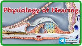
Cochlear System:
Cochlea consists of Scala Tympani, Scala Vestibuli and Scala Media.
When oval window is depressed, it created waves that travel through the cochlea. This will vibrate the basilar membrane and create neural activity through the spiral organ of corti.
Vestibulocochlear nerve:
Vestibular ganglion consists of superior and inferior division
Spiral (cochlear) ganglion located inside the cochlea. Several ganglions send out fibers that unite and form the cochlear division.
Vestibular and cochlear nerves unite and form vestibulocochlear nerve
They travel through the internal acoustic meatus.
Vestibular Nerve:
Vestibular nuclear complex (superior, lateral, medial and inferior)
VestibuloOcular Reflex (VOR)
○ Vestibuloocular reflex happens throuhg the medial longitudinal fasciculus
○ Fibers from medial vestibular nuclei to contralateral 6th cranial nerve, which sends fibers to contralateral third cranial nerve and fourth cranial nerve
VestibuloSpinal Pathway
○ Vestibulospinal reflex maintain posure and resist gravity
○ Lateral vestibular nuclei sends out lateral vestibulospinal tract
○ Medial vestibular nuclei sends out medial vestibulospinal tract that travel within the medial longitudinal fasciculus (MLF)
VestibuloCerebellar Pathway
○ Inferior vestibular nuclei sends out fibers that travel through the inferior cerebellar peduncle to the flocullonodular lobe and vermid. Fibers also come directly from the vestibulocochlear nerve
○ Cerebellum also sends fibers towards the vestibular nuclear complex to influence the vestibulospinal tract
VestibuloCerebral Pathway
○ Efferent vestibular projections to bilateral Ventral Posterior group of thalamus
○ Cortical regions of the brain known to be involved with vestibular processing:
○ Frontal eye fields: control eye movements and receive vestibular motion information
○ Primary somatosensory cortex (Areas 2v and 3a): map body location and movement signals
○ PIVC (ParietoInsular Vestibular Cortex): responds to body and head motion information
○ Posterior parietal cortex: motion perception and responds to both visual and vestibular motion cues
○ Hippocampus and parahippocampul regions: spatial orientation and navigation functions
Cochlear Nerve (Hearing pathway):
Synapse with ventral and dorsal cochlear nuclei.
Lateral lemniscus (lemniscus lateralis)
Crossing form trapezoid body.
Superior olivary complex (localizing the direction of sound)
Some synapse with trapezoid nucleus
Some synapse with nuclei of lateral lemniscus
Fibers go to Inferior colliculus, through inferior brachium to the medial geniculate body.
Most fibers reach medial geniculate body without relay in the inferior colliculus
Inferior colliculus controls a descending tract called Tectospinal tract that helps with the auditory reflex.
From medial geniculate body, auditory radiations goes to the primary auditory cortex.
Impulses also go to auditory association areas, Wernicke's area and Broca's area
Sources:
Singh, I. (2017). Human neuroanatomy (10th ed.).
Kozlowski, T. (2017). Memorix Anatomy: The Complete Study Guide. 2nd ed. Thieme Medical Publishers.
Neuroanatomy, Cranial Nerve 8 (Vestibulocochlear). Bruno Bordoni; Nicholas L. Mankowski; Daniel T. Daly.
Neuroanatomy, Auditory Pathway
Diana C. Peterson; Vamsi Reddy; Renee N. Hamel.
Pictures and visuals:
Complete Anatomy
Biorender
Powerpoint
Camtasia 2021