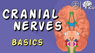
Cranial Nerves: Basic Anatomy Functions Effects of Damage and Clinical Tests Animation
00:00 The 12 pairs of cranial nerves (CN) overview
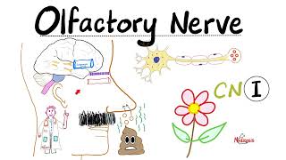
00:33 Olfactory nerve (CN 1)
00:55 Optic nerve (CN 2)
01:12 Oculomotor nerve (CN 3)
02:23 Trochlear nerve (CN 4)
02:56 Trigeminal nerve (CN 5)
04:09 Abducens nerve (CN 6)
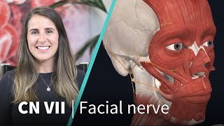
04:35 Facial nerve (CN 7)
05:36 Vestibulocochlear nerve (CN 8)
06:06 Glossopharyngeal nerve (CN 9)
07:03 Vagus nerve (CN 10)
08:20 Accessory nerve (CN 11)
09:07 Hypoglossal nerve (CN12)
Origin, termination, cranial exit/entrance, sensory/motor/parasympathetic functions, disorders (palsies), symptoms and evaluation.
Purchase a license to download a nonwatermarked version of this video on AlilaMedicalMedia(dot)com
Check out our new Alila Academy AlilaAcademy(dot)com complete video courses with quizzes, PDFs, and downloadable images.
©Alila Medical Media. All rights reserved.
Voice by : Marty Henne
All images/videos by Alila Medical Media are for information purposes ONLY and are NOT intended to replace professional medical advice, diagnosis or treatment.
Cranial nerves are numbered according to the order they exit the brain, from front to back.
Cranial nerve I olfactory nerve, is a sensory nerve responsible for the sense of smell.
Cranial nerve II optic nerve responsible for vision. It originates in the retina of the eye and ends in the thalamus. Optic nerve damage leads to partial or total blindness. Vision acuity is tested to assess nerve damage, one eye at a time.
Cranial nerve III oculomotor nerve controls most of the eye movements, as well as opening of eyelid and constriction of pupil. Origin = midbrain, contains both somatic and parasympathetic fibers. Somatic fibers innervate several extraocular, extrinsic eye muscles; while parasympathetic fibers terminate inside the eyeball and supply intrinsic eye muscles responsible for movement of the lens and pupil. Also for proprioception.
Oculomotor nerve palsy results in drooping eyelid, dilated pupil, loss of accommodation reflex, double vision, and inability to move eye in certain directions, characteristic sign = “down and out” deviation. Other tests include pupillary response to light, and ability to tract moving objects.
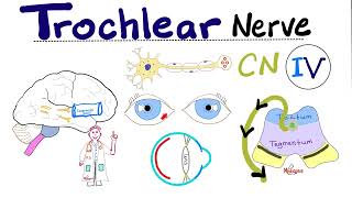
Cranial nerve IV trochlear nerve, originates in the midbrain and terminates in the superior oblique muscle of the eye. Damage to this nerve leads to double vision and eye deviation upward. The affected eye is unable to move down when looking to the direction of the normal eye.
Cranial nerve V trigeminal nerve has 3 divisions:
ophthalmic division: sensory information from the upper face.
maxillary division: sensory information from the middle section of the face,
mandibular division = mixed nerve: sensation from the lower face, including the anterior two thirds of the tongue, lower teeth and gums; motor muscles of mastication.
Cranial nerve VI abducens nerve, responsible for lateral eye movement. Origin: lower pons; termination: lateral rectus muscle of the eye.
Cranial nerves VII facial nerve, mixed nerve with diverse functions:
motor: muscles of facial expression,
sensory: taste sensations from the anterior twothirds of the tongue;
carries parasympathetic nerve impulses to tear glands and salivary glands.
Cranial nerve VIII vestibulocochlear nerve: vestibular nerve responsible for equilibrium, cochlear nerve responsible for hearing.
Cranial nerve IX glossopharyngeal nerve
sensory: from the upper pharynx, middle and outer ear, and the posterior third of the tongue, including taste buds.
visceral sensory signals from baroreceptors in the carotid sinus, and chemoreceptors in the carotid body, providing inputs for regulation of blood pressure and monitoring of blood oxygen,
parasympathetic innervation to the parotid salivary gland;
controls the stylopharyngeus muscle during speech, swallowing.
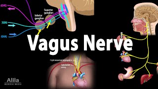
Cranial nerve X vagus nerve:
major parasympathetic nerve regulating pulmonary, cardiovascular and digestive activities;
controls most muscles of the pharynx, larynx, important role in swallowing and speech;
sensory information from the pharynx, larynx, and thoracic and abdominal areas, including baroreceptors and chemoreceptors in the aorta
Cranial nerve XI accessory nerve has both cranial and spinal roots. The spinal roots exit as the external branch and control the sternocleidomastoid and trapezius muscles.
Cranial nerve XII hypoglossal nerve controls extrinsic and intrinsic muscles of the tongue, responsible for various tongue movements and shapes required for normal swallowing and speech production.