Crohn Disease: Part 1 Active Inflammation

Audience: Radiologist, residents and fellows
Learning Objectives: At the end of this video the radiologist should be able to Classify the imaging findings of active inflammatory Crohn's disease of the small bowel.
Content:
Endoscopy is the gold standard for evaluation of mucosal disease.
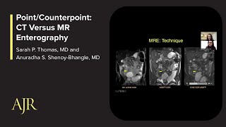
MR and CT Enterography are noninvasive diagnostic tools to assess stage severity, identify complications, evaluate treatment response, and assess for extraintestinal manifestations.
Standardized terminology is necessary to avoid confusion and miscommunication among radiologists and clinicians.
Consensus Guidelines on this terminology were published in 2018
Mural enhancement patterns:
Asymmetric Noncircumferential Mesenteric border
Stratified Secondary to submucosal edema, intramural fat, or inflammatory infiltration
Homogeneous / Symmetric Transmural hyperenhancement
Stricture Luminal narrowing in area of Crohn disease with unequivocal upstream dilation
Mild Upstream lumen 34 cm
Moderatetosevere Upstream lumen 4 cm or greater
Location and length described for potential surgical or endoscopic intervention
Determine if the stricture is due to active inflammation or fibrosis
Active inflammatory Crohn disease of the small bowel should be classified based on the degree of:
Wall thickening
Stricture
Upstream dilation
Mural hyperenhancement
Mural edema
Ulcerations
Bruining, David H., et al. "Consensus recommendations for evaluation, interpretation, and utilization of computed tomography and magnetic resonance enterography in patients with small bowel Crohn’s disease." Gastroenterology 154.4 (2018): 11721194.


![4 Common Foods that Make Crohn's Disease Worse [AVOID THIS]: Gut Health Expert](https://i.ytimg.com/vi/EV9pjSmDZ80/mqdefault.jpg)