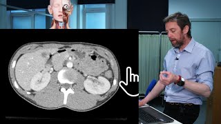
Cross sectional and imaging anatomy of the abdomen

This video deals with the anatomy of abdominal viscera and walls as they appear in transverse anatomical sections and axial CT sections. The video begins with a transverse anatomical section at the level of the T8 vertebra and continues down to the level of the L5 vertebra. There is a special emphasis on sections at the level of T10, T12, L1, L3, & L4.
00:04 Introduction
01:00 Section at the level of T8 vertebra
4:05 T10
8:30 T11/T12
12:45 T12
15:38 T12/L1
30:19 L1
32:29 L1/L2
36:25 L2/L3
39:35 L3
44:01 L4
The anatomical sections are arranged to match CT & MRI sections to provide a better understanding of the imaging anatomy of the abdomen. The arrangement of abdominal structures is followed in (22) serial transverse sections of the abdomen and compared to 5 representative axial CT sections at different levels.
The anatomical sections are selected from the Visible Human Project. For more information about this project refer to: http://www.nlm.nih.gov/research/visib...
Five short video clips are inserted where necessary to explain the 3D relations necessary to understand the 2D sections.
Presented and edited by Dr. Akram Jaffar (Ph.D.). Plastic model clips filmed by Parwiz Akbari (medical student). Filmed at the College of Medicine/ University of Sharjah, UAE. 2012.
This video and its channel are supported by the "Human Anatomy Education" page on Facebook / anatomyeducation
Related videos:
Crosssectional anatomy of the female pelvis and perineum • Cross sectional anatomy of the female...
Crosssectional and imaging anatomy of the thorax • Cross sectional and imaging anatomy o...
After completion of this video session, it is expected that you will be able to identify the approximate vertebral levels of transverse sections from T8L5 vertebrae. The following structures are identified in transverse anatomical sections or CT axial sections:
Vessels:
Inferior vena cava (IVC), aorta, esophagus, azygos vein, hepatic vein, portal vein, hepatic artery, splenic artery, splenic vein, celiac trunk, renal vein, superior mesenteric artery, superior mesenteric vein, common iliac artery, basivertebral vein.
Viscera:
Lungs, liver, spleen, gastroesophageal junction, fissure for ligamentum venosum, gallbladder, caudate lobe, porta hepatis, common hepatic duct, cystic duct; pancreas: head, neck, uncinate process, body, and tail; spleen, hepatic flexure, splenic flexure, kidney; duodenum: first, second, and third part; stomach, pyloric antrum, pyloric canal, pyloric sphincter, ascending colon, transverse colon, descending colon, cecum, jejunum, ileum, plicae circularis, rugae, umbilicus.
Musculoskeletal structure:
Costal origin of the diaphragm, crura of the diaphragm, rectus abdominis, external oblique, internal oblique, transverse abdominis, linea alba, serratus anterior, rectus sheath, latissimus dorsi, trapezius, erector spinae, posas major, quadrates lumborum, pectoralis major, thoracolumbar fascia, ilacus, gluteus medius, spinal cord, cauda equine, filum terminale, costovertebral joint, costotransverse joint, ribs, costal cartilages, intervertebral disc, sternum, iliac crest.
Other related videos
Coronal section 1 • sectional anatomy of the abdomen and ...
Coronal section 2 • sectional anatomy of the abdomen and ...
Coronal section 3 • Sectional anatomy of the abdomen and ...
Coronal section 4 • Sectional anatomy of the abdomen and ...
sagittal sections
• Sectional anatomy of the abdomen and ...
Related accounts
Twitter / akramjaffar
Facebook / anatomyeducation
SlideShare http://www.slideshare.net/AkramJaffar
LinkedIn / akramabo. .
Research gate https://www.researchgate.net/profile/...
Medtube https://medtube.net/users/akramjaffar
Instagram / akramjaffar
Academia https://dal.academia.edu/AkramJaffar