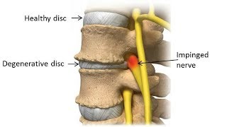
Degenerative disk disease progression over time
44 year old male Degenerative disk disease, disk herniation laminectomy and discectomy, progression over time after surgery. Weight gain result in venous obstruction and increase subcutaneus edema in the back.
MRI OF THE LUMBAR SPINE
HISTORY: Lumbar spine disc herniation. Low back pain.
TECHNIQUE: Multiplanar, multisequence MRI of the lumbar spine was performed on a 1.5 Tesla magnet.
COMPARISON: October 23, 2012, MR study of the lumbar spine.
FINDINGS: This dictation is based on the assumption that the patient has five lumbar type vertebral bodies.
Spinal alignment: 2 mm retrolisthesis of L5 on S1 is noted.
Bone marrow: There are Modic type I endplate changes at L5S1 level and L4L5 level, slightly more prominent as compared to October 23, 2012, at L4L5 level.
Paravertebral soft tissues: Unremarkable.
The conus terminates at the L1 level with normal morphology and signal intensity.
T12L1: Unremarkable.
L1L2: There is disc desiccation and a broad based 2 mm central disc protrusion.
L2L3: There is disc desiccation and a 3 mm broad based central disc protrusion, unchanged since the prior study.
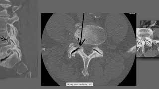
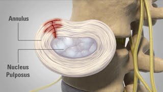
L3L4: There is disc desiccation and dorsal annular tear. Facet joint hypertrophy bilaterally is noted. There is no change since the prior study.
L4L5: There is disc desiccation and a 2 mm central disc protrusion. This is unchanged since the prior study. No spinal canal stenosis or nerve root compression.
L5S1: The patient is status post disc desiccation, disc space narrowing, and a broad based disc bulge. Facet joint hypertrophy bilaterally is noted. Left side laminotomy is seen, unchanged since the prior study. There is moderatetosevere left side neuroforaminal stenosis and moderate right side neuroforaminal stenosis, unchanged since the prior study. Compression on left L5 nerve root is possible.
IMPRESSION:
Status post left side L5 laminotomy and L5S1 diskectomy. There is multilevel degenerative disc disease and facet joint arthropathy with slight increase in segmental instability at L4L5 level since October 23, 2012, study. No evidence of spinal canal stenosis. Bilateral L5S1 neuroforaminal stenosis with probable left side nerve root compression are noted, unchanged since the prior study. No additional nerve root compression or spinal canal stenosis.