Digastric triangle : Boundaries and contents - Animated Gross anatomy head and neck

: / drgbhanuprakash
: https://t.me/bhanuprakashdr
: https://linktr.ee/DrGBhanuprakash
Digastric triangle
The digastric triangle is one of the paired triangles in the anterior triangle of the neck. The triangles of the neck are surgically focussed, first described from early dissectionbased anatomical studies which predated crosssectional anatomical description based on imaging (see deep spaces of the neck).
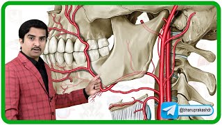
Boundaries
anteroinferiorly: anterior belly of digastric
posteroinferiorly: posterior belly of digastric
base: mandible
floor: mylohyoid, hyoglossus and middle pharyngeal constrictor
roof: skin, superficial fascia, platysma and deep fascia
Contents
anterior part of the triangle contains the submandibular gland
posterior part of the triangle contains the lower part of the parotid gland
facial artery is deep to the submandibular gland and the facial vein lies superficial to the gland
submandibular lymph nodes are situated near the gland
external carotid artery lies deep to the parotid gland before entering it. It is separated from the internal carotid artery by the styloglossus and stylopharyngeus muscles and the glossopharyngeal nerve
deep to the external carotid artery are the internal carotid artery, internal jugular vein and the vagus nerve
lying on the mylohyoid muscle are the submental and mylohyoid arteries and nerves
the hypoglossal and mylohyoid nerves are also found in the digastric triangle
#digastrictriangle #submandibulartriangle #anteriortriangleoftheneck #anatomy #headandneckanatomy #usmlestep1 #grossanatomydigastrictriangle