Dural Folds | Falx cerebri | Tentorium cerebelli | Falx cerebelli | Diaphragma sella | Attachments |

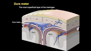
The dura mater is the outermost, thickest and toughest membrane covering the brain
There are two layers of dura:
a. An outer or endosteal layer which serves as an internal periosteum or endosteum or endocranium for the skull bones.
b. An inner or meningeal layer which surrounds the brain. The meningeal layer is continuous with the spinal dura mater.
The two layers are fused to each other at all places, except where the cranial venous sinuses are enclosed between them.
Endosteal Layer or Endocranium:
1. The endocranium is continuous:
a. With the periosteum lining the outside of the skull or pericranium through the sutures and foramina.
b. With the periosteal lining of the orbit through the superior orbital fissure.
2. It provides sheaths for the cranial nerves, the sheaths fuse with the epineurium outside the skull. Over the optic nerve, the dura forms a sheath which becomes continuous with the sclera.
3. Its outer surface is adherent to the inner surface of the cranial bones by a number of fine fibrous and vascular processes.
The adhesion is most marked at the sutures, on the base of the skull and around the foramen magnum.
Meningeal Layer
At places, the meningeal layer of dura mater is folded on itself to form partitions which divide the cranial cavity into compartments which lodge different parts of the brain.
The folds are:
1. Falx cerebri,
2. Tentorium cerebelli,
3. Falx cerebelli,
4. Diphragma sellae.
Falx cerebri
The falx cerebri is a large sickleshaped fold of dura mater occupying the median longitudinal fissure between the two cerebral hemispheres.
It has two ends:
1. The anterior end is narrow, and is attached to the crista galli.
2. The posterior end is broad, and is attached along the median plane to the upper surface of the tentorium cerebelli.
The falx cerebri has two margins:
1. The upper margin is convex and is attached to the lips of the sagittal sulcus.
2. The lower margin is concave and free.
The falx cerebri has right and left surfaces each of which is related to the medial surface of the corresponding cerebral hemisphere.
Three important venous sinuses are present
in relation to this fold.
1. The superior sagittal sinus lies along the upper margin;
2. The inferior sagittal sinus along the lower margin
3. The straight sinus along the line of attachment of the falx to the tentorium cerebelli
Tentorium cerebelli
• The tentorium cerebelli is a tentshaped fold of dura mater, forming the roof of the posterior cranial fossa.
• It separates the cerebellum from the occipital lobes of the cerebrum,
• broadly divides the cranial cavity into supratentorial and
infratentorial compartments.
• The infratentorial compartment, in other words, is the posterior cranial fossa containing the hindbrain and the lower part of the midbrain.
• The tentorium cerebelli has
• a free margin and
• an attached margin
• The anterior free margin is Ushaped and free.
• The ends of the 'U' are attached anteriorly to the anterior clinoid
processes.
• This margin bounds the tentorial notch which is occupied by the midbrain and the anterior part of the superior vermis.
• The outer or attached margin is convex.
• Posterolaterally, it is attached to the lips of the transverse sulci
on the occipital bone, and on the posteroinferior angle of the parietal bone.
• Anterolaterally, it is attached to the superior border of the
petrous temporal bone and to the posterior clinoid processes.
• Along the attached margin, there are the transverse and superior
petrosal venous sinuses.
The trigeminal or Meckel's cave is a recess of dura mater present in relation to the attached margin of the tentorium.
It is formed by evagination of the inferior layer of the tentorium over the trigeminal impression on the petrous temporal bone.
It contains the trigeminal ganglion.
The free and attached margins of the tentorium cerebelli cross each other near the apex of the petrous temporal bone. Anterior to the point of crossing, there is a triangular area which forms the posterior part of the roof of the cavernous sinus, and is pierced by the third and fourth cranial nerves. The tentorium cerebelli has two surfaces. The superior surface is convex and slopes to either side from the median plane.
The falx cerebri is attached to this surface, in the midline; the straight sinus lies along the line of this attachment. The superior surface is related to the occipital lobes of the cerebrum.
The inferior surface is concave and fits the convex superior surface of the cerebellum. The falx cerebelli is attached to its posterior part
Falx cerebelli
The falx cerebelli is a small sickleshaped fold of dura mater projecting forwards into the posterior cerebellar notch. The base of the sickle is attached to the posterior part of the inferior surface of the tentorium cerebelli in the median plane.
Follow me in blogspot https://humananatomylessons.blogspot...


![Neck Dissection Surgical Anatomy: OR FAQs & Answers [201] Didactic](https://i.ytimg.com/vi/oSQLNfo4rXs/mqdefault.jpg)