Embryology of the Face (Easy to Understand)

The development of the face explained in a very simple way.
This is part one of two, in the next videos I will discuss the embryology of the teeth and eyes.
If you are completely new to embryology and you want to understand it quickly, this should be the first video you watch:
• Introduction to Embryology Fertilis...
Recommended Text
Easy Embryology is a book that is dedicated to the simplification of embryology. It is available at https://drminass.com/product/easyembr.... Contact Dr. Minass for more information.
Interact With Dr. Minass!
Website https://www.drminass.com
Email [email protected]
Patreon / drminass
Facebook / m1na55
Instagram @m1.nass
Post Address to:
Minass
Parcel Locker 10106 04448
59 Penshurst Street
Willoughby, NSW
Australia 2068
If you would like to support the channel you can donate here:
https://www.paypal.com/donate/?token=...
SUMMARY FOR YOUR NOTES:
How the face develops:
At the end of the forth week, facial prominences consisting primarily of neural crestderived mesenchyme and formed mainly by the first pair of pharyngeal arches appear.
Maxillary prominences are lateral to the stomoduem
Mandibular prominences are caudal.
Frontonasal prominence is formed by proliferation of mesenchyme.
During the fifth week, the nasal placodes invaginate to form nasal pits.
During weeks 57 the maxillary prominences continue to grow, filling the medial part of the face, compressing the medial nasal prominences. The cleft between the medial nasal prominence and the maxillary prominence is lost, and the two fuse.
The upper lip formed by the two medial nasal prominences and the two maxillary prominences.
Lower lip and jaw form from the mandibular prominence that merge across the midline. Initially the maxillary and lateral nasal prominences are separated by a deep furrow, the nasolacrimal groove. Ectoderm in the floor of this groove forms a solid epithelial cord that detaches from the overlying ectoderm.
After canalisation, the cord forms the nasolacrimal duct; its upper end widens to form the lacrimal sac.
The nasolacrimal duct runs from the medial corner of the eye to the inferior meatus of the nasal cavity, and the maxillary prominences enlarge to form the cheeks and maxillae.
The nose is formed from the five facial prominences: the frontal prominence gives rise to the bridge, the merged medial nasal prominence provide the crest and tip and the lateral nasal prominence form the sides.
Intermaxillary segment (the primary palate)
The result of medial growth of the two MNP merge not only at the surface but deeper. The structure formed is the intermaxillary segment:
1. Labial component which forms the philtrum of the upper lip
2. Upper jaw components which carries the four incisor teeth
3. A palatal component, which forms the triangular primary palate
The intermaxillary segment is continuous with the rostral portion of the nasal septum which is formed by the frontal prominence
The secondary palate:
Outgrowths called the palatine shelves from the maxillary process appear during week 6.
They grow obliquely and inferiorly but end up in their horizontal position above the tongue finally during week 78.
The outgrowths fuse with the anterior plate from the intermaxillary segment.
The incisive foramen marks the midline between the primary and secondary palates.
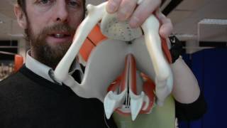
Nasal cavities:
During week 6 nasal pits deepen.
Oronasal membrane degenerates
The definitive choanae lies at the junction of the nasal cavity and the pharynx.
The paranasal sinuses develop as diverticula of the lateral nasal wall and extends into the maxilla, ethmoid, frontal, and sphenoid bones.
They reach their full size by puberty and they contribute to the shape of the face.