Embryology of the Female Reproductive System I (Easy to Understand)

The development of the female reproductive system explained in a very simple way.
If you are completely new to embryology and you want to understand it quickly, this should be the first video you watch:
• Introduction to Embryology Fertilis...
Recommended Text
Easy Embryology is a book that is dedicated to the simplification of embryology. It is available at https://drminass.com/product/easyembr.... Contact Dr. Minass for more information.
Interact With Dr. Minass!
Website https://www.drminass.com
Email [email protected]
Patreon / drminass
Facebook / m1na55
Instagram @m1.nass
Post Address to:
Minass
Parcel Locker 10106 04448
59 Penshurst Street
Willoughby, NSW
Australia 2068
SUMMARY FOR YOUR NOTES:
The key to sexual dimorphism is the Y chromosome which contains the testis determining gene called the SRY (sex determining region on Y) gene
The SRY protein is the testis determining factor and under its influence male development occurs
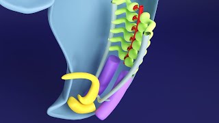
Gonads
The sex of the embryo is determined at the time of fertilisation, but the gonads do not acquire male or female morphological characteristics until the seventh week of development
The gonads appear initially as a pair of longitudinal ridges called genital ridges
Genital ridges are formed by proliferation of the epithelium and a condensation of underlying mesenchyme
Germ cells appear in the 6th week of development
Primordial germ cells originate in the epiblast, migrate through the primitive streak, and by the third week reside among endoderm cells in the wall of the yolk sac close to the allantois
During the 4th week they migrate by ameboid movement along the dorsal mesentery of the hindgut, arriving at the primitive gonads at the beginning of the 5th week, invading the genital ridges
While waiting for the germ cells to arrive, the genital ridge epithelium proliferates and penetrate the underlying mesenchyme where they form cords called the primitive sex cords.
Ovary
In females with a XX chromosome and no Y chromosome, primitive sex cords differentiate into irregular cell clusters.
These clusters containing primitive germ cells are the medullary part of the ovary. Later they disappear and are replaced by a vascular stroma that forms the ovarian medulla.
The surface epithelium of the female gonad, unlike that of the male, continues to proliferate.
In the 7th week, it gives rise to a second generation of cords, cortical cords, which penetrate the underlying mesenchyme but remain close to the surface.
In the third month, these cords split into isolated cell clusters which continue to proliferate and begin to surround each oogonium with a layer of epithelial cells called follicular cells.
Together the oogonia and follicular cells constitute a primordial follicle
Genital ducts
Initially both male and female embryos have 2 pairs of genital ducts: mesonephric (Wolffian) ducts and paramesonephric (Mullerian) ducts
The paramesonephric duct arises as a longitudinal growth of the epithelium on the anterolateral surface of the urogenital ridge
Cranially the duct opens into the abdominal cavity with a funnel like structure
Caudally it first runs lateral to the mesonephric duct from the opposite side
The two ducts are initially separated by a septum but later fuse to form the uterine canal.
The caudal tip of the combined ducts projects into the posterior wall of the urogenital sinus where it causes a small swelling, the paramesonephric tubercle.
The mesonephric ducts open into the urogenital sinus on either side of the Mullerian tubercle.
The paramesonephric ducts develop into the main genital ducts of the female
With descent of the ovary, first 2 parts develop into the uterine tube
The caudal parts fuse to form the uterine canal.
After the ducts fuse in the midline, a broad transverse pelvic fold is established (broad ligament of uterus)
The fused paramesonephric ducts give rise to the corpus and cervix of uterus
They are surrounded by a layer of mesenchyme that forms the muscular coat of the uterus, the myometrium and its peritoneal covering (perimetrium)
V (YouTube won't allow for the full word to be used here .)
The solid tip of the paramesonephric ducts reaches the urogenital sinus and two solid parts grow out from the pelvic part of the sinus (sinoV bulbs) which proliferate and form a solid V plate.
By the 5th month the V outgrowth is entirely canalised
the expansions of the V around the cervix, fornicies, are paramesonephric origin.
Therefore the V has a dual origin ,the upper portion derived from uterine canal and lower portion derived from the urogenital sinus
The lumen of the V remains separated from that of the urogenital sinus by a thin tissue called the hymen which consists of epithelial lining of the sinus and a thin layer of V cells. It usually develops a small opening during perinatal life.