Hip Pain Strain Of The Rectus Femoris anatomy - Everything You Need To Know - Dr. Nabil Ebraheim

Dr. Ebraheim’s educational animated video illustrates the condition of Rectus Femoris pain / strain and its associated anatomy.
This animated video explains the anatomy of the Rectus Femoris muscle and hip pain that is caused by strain of the rectus femoris muscle. This condition usually occurs in active individuals and runners. The action of the rectus femoris muscle is explained. Palpation of the rectus femoris muscle and contraction test of the rectus femoris muscle is explained. Injection of the rectus femoris is mentioned. Palpation of the rectus femoris is outlined. The treatment of rectus femoris tear is outlined, stretching exercise and active release technique.
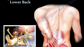
Hip Pain is strain of the rectus femoris and its anatomy.
One of the causes of hip pain that is usually forgotten is a strain of the rectus femoris muscle and tendon.
It causes groin pain or anterior hip pain in some athletes.
Who are these athletes that gets problems from rectus femoris strain?
Runners.
Baseball playoffs.
Hocky players.
Soccer players.
It will be sudden like kicking the ball in soccer or sprinting from a standing position.
It’s an overuse problem with repeated tearing and repeated stress and stretching of the tendon.
There will be some scarring, adhesion, or tightness.
Strain of the rectus femoris is similar to a tennis elbow type of problem or Achilles tendonitis.
It is important to know that there is a lot of causes of hip pain, especially the anterior hip pain, it can be something inside the joint:
• Loose body.
• liberal tear
• Avascular necrosis of the femoral head.
• Arthritis.
Or something outside the joint:
• The illiopsoas bursitis.
When you work the patient up for hip pain, make sure to include the rectus femoris muscle strain, which occur from tearing of the muscle fibers due to stretching of this muscle.
The rectus femoris is the anterior muscle of the quadriceps femoris.
The quadriceps femoris is a group of four muscles on the anterior thigh.
The rectus femoris has two heads, originating from the pelvis:
1 The straight head arises from the anterior inferior iliac spine.
2 The reflected head arises from the groove superior to the acetabulum.
The rectus femoris muscle is inserted to the superior border if the patella through the common quadriceps tendon.
This muscle flexes the thigh at the hip and it extends the knee.
The innervation comes from the femoral nerve: L2, L3, and L4.
When the muscle crosses the hip anteriorly, it flexes the hip.
Tears or strains of the rectus femoris muscle can be an acute process from forcible acentric contraction of the muscle.
These injuries are usually more distal on the thigh or near the knee.
Patient with a strain will have pain in the groin area or anterior part of the hip, most commonly you will find that the pain is more distal.
The chronic overuse injury will give you anterior hip pain, usually near the anterior inferior iliac spine.
In adolescents, you will have avulsion of the anterior inferior iliac spine.
And the treatment is no surgery, just some rest and crutches.
Palpate the site of the pain.
Go around the hip region and palpate the anterior superior iliac spine.
The ASIS is where the Sartorius is attached too.
Immidiatly distal to that you find the soft spot or soft groove followed by the anterior inferior iliac spine, you try to palpate carefully because It maybe the area of the rectus femoris sprain if this area is painful.
Also you want to palpate the tensor fascia Lata and the iliotibial band around the hip area.
Try to palpate the rectus femoris muscle through its entire length because you may find pain in the midthigh or distally (common).
Then you will do the provocative test: resisted hip flexion will produce pain.
This situation is different then pulled groin which would be abductor muscle strain and the patient would have pain and decreased strength with resisted leg abduction, and it's called compression contraction test.
So we finish the exam for the rectus femoris muscle strain then we will probably not need any xrays, but in adolescents we may get an xray to make sure that there is no avulsion fracture of the anterior inferior iliac spine.
If you get an Xray probably it will be normal.
Get an MRI I tough cases that are not getting better with treatment.
Treatment is done by:
Ice
Rest
NSAIDS
Physical therapy: stretching and strengthening, active release techniques.
Injection
Surgery is really done.
Follow me on twitter:
https://twitter.com/#!/DrEbraheim_UTMC
Donate to the University of Toledo Foundation Department of Orthopaedic Surgery Endowed Chair Fund:
https://www.utfoundation.org/foundati...