Histology Of Retina

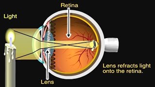
The retina (Latin: rēte) is the innermost, lightsensitive layer of tissue of the eye of most vertebrates and some molluscs. The optics of the eye create a focused twodimensional image of the visual world on the retina, which translates that image into electrical neural impulses to the brain to create visual perception. The retina serves a function analogous to that of the film or image sensor in a camera.
The neural retina consists of several layers of neurons interconnected by synapses, and is supported by an outer layer of pigmented epithelial cells. The primary lightsensing cells in the retina are the photoreceptor cells, which are of two types: rods and cones. Rods function mainly in dim light and provide blackandwhite vision. Cones function in welllit conditions and are responsible for the perception of colour, as well as highacuity vision used for tasks such as reading. A third type of lightsensing cell, the photosensitive ganglion cell, is important for entrainment of circadian rhythms and reflexive responses such as the pupillary light reflexRetinal layers
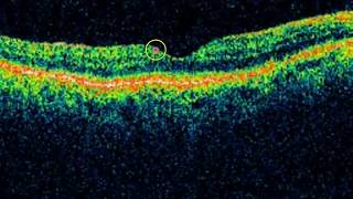
Section of retina
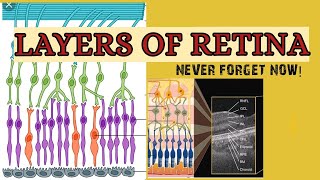
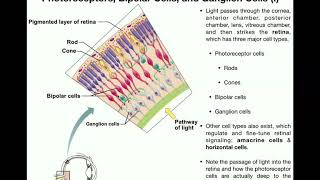
Rods, cones and nerve layers in the retina. The front (anterior) of the eye is on the left. Light (from the left) passes through several transparent nerve layers to reach the rods and cones (far right). A chemical change in the rods and cones send a signal back to the nerves. The signal goes first to the bipolar and horizontal cells (yellow layer), then to the amacrine cells and ganglion cells (purple layer), then to the optic nerve fibres. The signals are processed in these layers. First, the signals start as raw outputs of points in the rod and cone cells. Then the nerve layers identify simple shapes, such as bright points surrounded by dark points, edges, and movement.
Illustration of the distribution of cone cells in the fovea of an individual with normal colour vision (left), and a colourblind (protanopic) retina. Note that the center of the fovea holds very few bluesensitive cones.
Distribution of rods and cones along a line passing through the fovea and the blind spot of a human eye
The vertebrate retina has ten distinct layers. From closest to farthest from the vitreous body:
Inner limiting membrane – basement membrane elaborated by Müller cells.
Nerve fibre layer – axons of the ganglion cell bodies (note that a thin layer of Müller cell footplates exists between this layer and the inner limiting membrane).
Ganglion cell layer – contains nuclei of ganglion cells, the axons of which become the optic nerve fibres, and some displaced amacrine cells.
Inner plexiform layer – contains the synapse between the bipolar cell axons and the dendrites of the ganglion and amacrine cells.
Inner nuclear layer – contains the nuclei and surrounding cell bodies (perikarya) of the amacrine cells, bipolar cells, and horizontal cells.
Outer plexiform layer – projections of rods and cones ending in the rod spherule and cone pedicle, respectively. These make synapses with dendrites of bipolar cells and horizontal cells. In the macular region, this is known as the Fiber layer of Henle.
Outer nuclear layer – cell bodies of rods and cones.
External limiting membrane – layer that separates the inner segment portions of the photoreceptors from their cell nuclei.
Inner segment / outer segment layer – inner segments and outer segments of rods and cones. The outer segments contain a highly specialized lightsensing apparatus.
Retinal pigment epithelium – single layer of cuboidal epithelial cells (with extrusions not shown in diagram). This layer is closest to the choroid, and provides nourishment and supportive functions to the neural retina, The black pigment melanin in the pigment layer prevents light reflection throughout the globe of the eyeball; this is extremely important for clear vision.
These layers can be grouped into 4 main processing stages: photoreception; transmission to bipolar cells; transmission to ganglion cells, which also contain photoreceptors, the photosensitive ganglion cells; and transmission along the optic nerve. At each synaptic stage there are also laterally connecting horizontal and amacrine cells.
The optic nerve is a central tract of many axons of ganglion cells connecting primarily to the lateral geniculate body, a visual relay station in the diencephalon (the rear of the forebrain). It also projects to the superior colliculus, the suprachiasmatic nucleus, and the nucleus of the optic tract. It passes through the other layers, creating the optic disc in primates.


![Histology of the Retina [Special Senses Histology Part 3 of 4]](https://i.ytimg.com/vi/zG4ZhMusF4Y/mqdefault.jpg)