Hyoid bone osteology - Gross anatomy Attachments Movements Clinical significance - USMLE

: / drgbhanuprakash
: https://t.me/bhanuprakashdr
: https://linktr.ee/DrGBhanuprakash
Hyoid bone , osteology USMLE Step 1 : Medvizz anatomy animated lectures
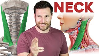
Hyoid bone
With the exception of the cervical vertebrae, the hyoid bone is the only bone located in the anterior neck. Unlike other bony structures, the hyoid bone does not directly articulate with other bones. Instead, it is connected to neighbouring bones by muscular and ligamentous attachments.
Muscles that insert on the upper surface of the bone are known as suprahyoid muscles, while those attached to the lower surface are infrahyoid muscles. This article will explore the embryology, anatomy and muscular attachments of this structure.
Gross anatomy
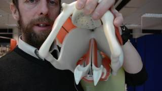
The hyoid bone is a Ushaped bone that is held in place by the strap muscles of the anterior triangle of the neck. The bone has a central body (forming the center of the “U”) with two smaller protruding structures on the superior surface (lesser horns) and two larger bony protrusions from the body (greater horns).
The body is quadrilateral, laterally stretched and irregular in shape. It forms the convexity of its classical Ushape, with its outer (anterior) border forming the outer convexity and the inner (posterior) border forming the concavity. There is a vertical median ridge located in the midline of the body that rarely projects to the lower surface. The body is situated at an oblique angle with the anterior end pointing superiorly and the posterior end pointing inferiorly. Immediately posterior to the body of the hyoid bone is a bursa, thyrohyoid membrane and areolar tissue that separates the hyoid bone from the epiglottis.
The greater cornua (horns) extend from the lateral extremity of the body in a posterolateral direction. They are wider proximally and become increasingly narrow until they terminate in tubercles. They are also flattened horizontally.
At the junction of the body and each greater cornu is a conical protruding bone known as the lesser cornua (horns). Fibrous tissue connects the lesser cornua to the body of the hyoid bone. There are also instances where each lesser cornu articulates with the ipsilateral greater cornu by way of a synovial joint that ossifies in later decades of life.
Attachments
The hyoid bone has superior and inferior muscular attachments that keep it in place in the anterior region of the neck. Based on this, the muscles attaching to the hyoid bone are divided into the suprahyoid and infrahyoid muscles.
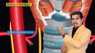
There are also one membranous and two ligamentous attachments to the bone. The thyrohyoid membrane is an extrinsic membrane of the larynx that connects the superior border of the thyroid lamina and its superior horns to the superior aspect of the greater cornua and body of the hyoid bone. It accesses the superior surface of the hyoid bone by passing behind the bone and the bursa.
The thyrohyoid membrane functions as the lateral walls of the piriform recess. The internal laryngeal nerve, along with the superior laryngeal arteries and veins, also pierce the membrane. Its primary responsibility is anchoring the laryngeal skeleton to the hyoid bone.
The hyoepiglottic ligaments are one of several extrinsic ligaments of the larynx. The bilateral ligaments are collections of fibrous tissue that are responsible for attaching the anterior and lateral aspects of the epiglottis to the body and greater cornu of the hyoid bone. Consequently, they give rise to the glossoepiglottic folds that surround the valleculae (depressions at the root of the tongue).
The stylohyoid ligament commences at the apex of the styloid process and inserts in the lesser cornu of the hyoid bone. It is crossed inferiorly by hyoglossus. The fibrous ligament, which is closely related to the lateral oropharyngeal wall, provides a point of attachment for the middle pharyngeal constrictors and some fibers of styloglossus. The ligament is a derivative of the second branchial arch and may be calcified.
#hyoidbone #hyoidboneanatomy #hyoidboneanimation #hyoidboneosteology #drgbhanuprakash #anatomy #anatomyanimations #anatomyusmle #anatomymbbs #anatomyvideos #anatomylectures