YouTube doesn't want you know this subscribers secret

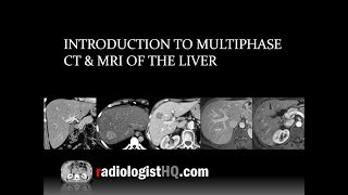
HypoVascular Masses of the Liver

Audience: Radiology Residents, Fellows and Attendings
Learning Objectives:
Identify the distinguishing features of the most common hypervascular and hypovascular masses of the liver
Compare and contrast common benign and malignant liver lesions
Recognize exceptions and atypical presentations of lesions, particularily with Eovist/Primovist
Summary:
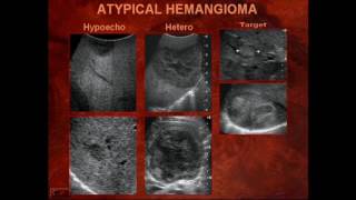
Hemangioma
T1 hypointense, T2 moderately hyperintense
Peripheral nodular discontinuous arterial phase hyperenhancement
Progressive enhancement following blood pool
Cholangiocarcinoma/adenocarcinoma metastasis
T1 hypointense, T2 mildy to moderately hyperintense
Rim arterial phase hyperenhancement (or arterial phase hypoenhancement)
Progressive enhancement not following blood pool


![CT Differential Diagnosis of Focal Hepatic Lesions [Basic Radiology]](https://i.ytimg.com/vi/PxSRWNqnDf4/mqdefault.jpg)
Recommended