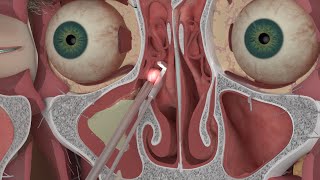
Imaging Anatomy of the Paranasal Sinuses

Anatomy of the paranasal sinuses on imaging. In this video we'll explore the anatomy of the paranasal sinuses on CT. A good understanding of paranasal sinus anatomy is imperative to understand imaging patterns in rhinosinusitis. Imaging anatomy is complicated by the presence of many anatomical variants. A good understanding of paranasal sinus anatomy is needed to detect predisposing factors for the development of (chronic) sinusitis and to identify possible surgical risks. This presentation was made by dr. Simon Nicolay (UZ Antwerp, Belgium) and dr. Sven Dekeyzer (UZ Ghent, Belgium).
0:00 Introduction + topics
1:26 General sinonasal anatomy
3:50 The nasal cavity
6:29 The nasal septum
9:05 Function of the nasal cavity
10:54 The nasal turbinates
14:42 The nasal meatus
15:35 Function of the paranasal sinuses
18:41 Drainage pathways of the paranasal sinuses
19:34 The sphenoethmoidal recess
21:48 The frontal recess
22:46 The ethmoid bulla
23:44 The (ethmoidal) infundibulum
24:07 The ostiomeatal complex
26:08 The nasolacrimal system
27:31 The infraorbital canal and supraorbital notch
28:19 The anterior and posterior superior alveolar canals
29:19 Anatomic variants
30:21 Nasal cavity variants
30:37 Septal deviation
33:32 Septal defect
35:09 Concha bullosa
37:12 Paradoxical middle turbinate
38:24 Olfactory Fossa
39:30 Keros classification
42:14 Sphenoid sinus variants
43:02 Sphenoid sinus pneumatization
44:22 Sphenoid skull base pneumatization
47:15 Vidian canal protrusion / dehiscence
48:18 Optic nerve and carotid canal protrusion / dehiscence
49:41 Sinus septum insertion on the carotid canal
51:27 Ehtmoid cell variants
51:58 Ethmoid bulla
53:08 AggerNasi cell
53:45 Frontal recess cells
55:14 Haller cells
56:18 Supraorbital air cells
1:00:20 Onodi cells
1:03:40 Lamina papyracea
1:04:17 Adherent uncinate process
1:05:24 Key Messages
1:06:24 Anatomic variants that (might) narrow the sinonasal outflow tracts
1:06:54 Anatomic variants that (might) pose surgical risks
1:10:07 References and word of thanks to dr. Simon Nicolay
This video is brought to you by the neuroradiologist:
/ theneuroradguy
/ the_neurora. .
https://theneuroradiologist.org/
#radiology #neuroradiology #neurology #medicalstudent #neuroradiologist #theneuroradiologist #otorhinolaryngology #anatomy #MRI #medical #sinuses


![The moment we stopped understanding AI [AlexNet]](https://i.ytimg.com/vi/UZDiGooFs54/mqdefault.jpg)