Jejunum: Gross anatomy Blood supply Venous drainage Nerve supply and Histology | USMLE Step 1

: / drgbhanuprakash
: https://t.me/bhanuprakashdr
: https://linktr.ee/DrGBhanuprakash
Gross Anatomy of Jejunum
Position and Size
The jejunum is the middle part of the small intestine, lying between the duodenum and ileum.
It typically begins in the left upper quadrant of the abdomen and occupies the umbilical region, extending in numerous loops.
It measures approximately 2.5 meters in length, though this can vary.
Vascular Supply to the Jejunum
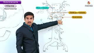
Arterial Supply
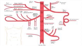
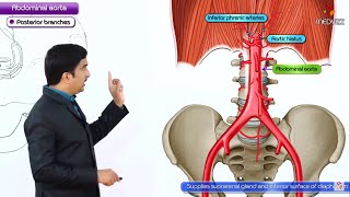
The superior mesenteric artery (SMA), a branch of the abdominal aorta, provides the primary arterial blood supply to the jejunum.
The SMA gives off several jejunal branches, which further divide into arcades.
These arcades then give rise to straight vessels, known as vasa recta, that supply the jejunum.
Venous Drainage
The venous drainage of the jejunum mirrors its arterial supply.
The jejunal veins, which follow the path of the arteries, drain into the superior mesenteric vein (SMV).
The SMV ultimately drains into the hepatic portal vein, leading to the liver.
Lymphatic Drainage of Jejunum
Lymph from the jejunum drains into the mesenteric lymph nodes first.
These nodes ultimately drain into the superior mesenteric lymph nodes.
Finally, the lymph drains into the cisterna chyli, a significant collection point for intestinal lymph.
Nerve Supply of Jejunum
Innervation
The nerve supply to the jejunum is primarily via the superior mesenteric plexus.
This plexus receives fibers from both the sympathetic and parasympathetic divisions of the autonomic nervous system.
Sympathetic fibers originate from the lesser splanchnic nerves and parasympathetic fibers from the vagus nerve.
Histology of Jejunum
Histological Layers
The wall of the jejunum, like the rest of the gastrointestinal tract, consists of several layers: mucosa, submucosa, muscularis propria, and serosa.
The mucosa is characterized by large, closely packed circular folds known as plicae circulares and villi, which increase the surface area for absorption.
The mucosa contains simple columnar epithelial cells, including goblet cells (for mucus secretion) and Paneth cells (for antimicrobial secretion).
Brunner's glands, found in the submucosa, secrete an alkaline fluid composed of mucus, which helps neutralize gastric acid in the chyme.
The muscularis propria consists of two layers of smooth muscle: inner circular and outer longitudinal.
The serosa, the outermost layer, is composed of mesothelial cells.
Distinguishing Features
Compared to the ileum, its distal counterpart, the jejunum has fewer Peyer's patches (aggregated lymphoid nodules), more prominent plicae circulares, and more extended vasa recta. These features are crucial for differentiating jejunum from ileum in histological identification.
#jejunum #jejunumanatomy #smallintestine #fmge #fmgevideos #rapidrevisionfmge #fmgejan2023 #mbbslectures #nationalexitexam #nationalexittest #neetpg #usmlepreparation #usmlestep1 #fmge #usmle #drgbhanuprakash #medicalstudents #medicalstudent #medicalcollege #neetpg2023 #usmleprep #usmlevideos #usmlestep1videos #medicalstudents #neetpgvideos #abdomenanatomy #usmleanatomy