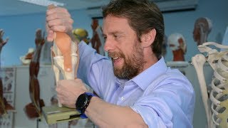
LIGAMENTS OF THE KNEE

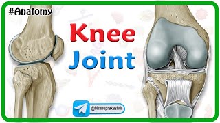
The knee is the largest joint in the human body. It is a compound joint. Not only is it where the femur, or thigh bone, meets the tibia, or shin bone – at the tibiofemoral joint, but it is also where the femur meets the patella, or kneecap – at the patellofemoral joint.
There are several ligaments around the knee joint and these are crucial because they limit movements and stabilize the joint. Ligaments are durable bands of fibrous tissue that connect joints and strengthen them.
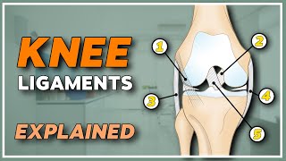
There are two main pairs of ligaments in the knee – the cruciate ligaments, which are inside your knee joint, and the collateral ligaments, which run on either side of the knee.
The cruciate ligaments can be seen through the intercondylar notch of the femur. There’s the anterior cruciate ligament, or ACL, and the posterior cruciate ligament, or PCL, which stabilize the knee. The ACL prevents the tibia from being pushed forward relative to the femur, while the PCL prevents it from being pushed backwards relative to the femur.
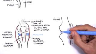
Now for the collateral ligaments – there’s a medial collateral ligament and a lateral collateral ligament. Medial means at the middle, while lateral means on the side. Hence, the medial collateral ligament is found on the inner side of the knee, running from the femur to the tibia. The lateral collateral ligament is found on the outer side of the knee, however, it runs from the femur to the fibula! Note that the medial collateral ligament is significantly wider than the lateral collateral ligament. Together, the medial and lateral collateral ligaments resist sideways movements of the bones relative to one another.
Looking back inside the intercondylar notch, we can see the transverse ligament – otherwise called the anterior meniscomeniscal ligament. This ligament connects the anterior lateral meniscus to the anterior medial meniscus. This ligament is important during knee extension, since it prevents the anterior horns of the menisci from coming forward, which would cause the condyles of the femur and tibia to put pressure on them.
A ligament discovered in 2013 is the anterolateral ligament, or ALL. It originates at the femur and inserts into the tibia. It is thought that it might control internal tibial rotation.
Here we have the ligament of Wrisberg, also called the posterior meniscofemoral ligament. This ligament attaches to the posterior lateral meniscus and crosses behind the PCL to attach to the medial condyle of the femur.
Now I’d like to bring your attention to a structure that some call a tendon, and some call a ligament. As a reminder, the difference between ligaments and tendons is that ligaments connect bones to bones, while tendons connect bones to muscles. Here we have the patellar tendon… which some people call the patellar ligament. The patellar tendon connects the patella (or kneecap) to the tibia. Since these are two bones, shouldn’t it be a ligament? Well, this structure is really connecting the quadriceps muscle to the tibia. The patellar tendon is part of a more extensive mechanism, which includes the tibia, the patellar tendon, the patella, the quadriceps tendon, and the quadriceps muscle. Together, these structures allow you to straighten your knee. As a side note, the patella is what is known as a floating sesamoid bone. A sesamoid is a bone embedded in a tendon.
To close off, I just want to bring your attention to a couple of other structures visible in this model. The medial and lateral menisci are composed of connective tissue with extensive collagen fibers. They protect the ends of the bones from rubbing against each other. Similarly to the collateral ligaments, the medial one is on the inner side of your knee and the lateral one is on the outer side of your knee.
We can also see the articular cartilage on this model. This smooth, white tissue covers the ends of bones where they converge at joints, minimizing friction and allowing bones to glide over each other. As you get older, the articular cartilage and the menisci wear down, exposing underlying bone. This changes the load distribution and biomechanics of your knee and causes pain and inflammation as your bones grind together.
3D model modified from https://www.turbosquid.com/FullPrevie...