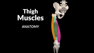
Muscles of the Leg (Division Origin Insertion Functions)

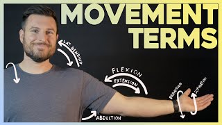
The term ''Foot Extension'' means ''Dorsiflexion'' (To avoid confusion)
Content
Introduction 0:00
Division of the Leg Muscles 0:20
Anterior Group 00:40
Extensor Hallucis Longus 1:17
Extensor Digitorum Longus 1:39
Tibialis Anterior 2:03
Lateral Group 2:30
Fibularis Brevis 2:53
Fibularis Longus 3:09
Posterior Group Deep 3:43
Popliteus 4:12
Tibialis Posterior 4:31
Flexor Digitorum Longus 5:00
Flexor Hallucis Longus 5:21
Posterior Superficial Layer 5:42
Triceps Surae 5:46
Plantaris 6:47
Channel membership: / @taimtalksmed
Follow my IG: / taimtalksmed
Donation link: https://www.buymeacoffee.com/taimtalk...
Muscles of the Leg
Anterior Group [3]t
Lateral Group [2]
Posterior Group [6] – Deep + Superficial Layers
Anterior Group [3]
Extensors of the Leg
Tendons pass under the extensor retinaculum
Innervated by the deep fibular nerve
Extensor Hallucis Longus
musculus extensor hallucis longus
O:
Medial Surface of Fibula
Interosseous membrane
I: Distal Phalanx of big toe
phalanx distalis hallucis
F:
Extension of big toe
Extension of Foot
Supination + adduction of Foot
Extensor Digitorum Longus
musculus extensor digitorum longus
O:
Lateral Condyle of Tibia
Fibula
Interosseous membrane
I:
Middle and Distal Phalanx of 2nd – 5th toe
5th metatarsal bone
F:
Extension of 2nd – 5th toe
Extension of Foot
Tibialis Anterior
musculus tibialis anterior
O:
Lateral Condyle of Tibia
Lateral Surface of Tibia
Interosseous membrane
I:
Base of the 1st Metatarsal Bone
Medial Cuneiform
F:
Extension of Foot
Supination + Adduction of Foot
Lateral Group [2]
Consist of two fibular muscles that originate on the lateral surface of fibula
Both muscles run behind the lateral malleolus under the superior and inferior fibular retinaculum
Innervated by the superficial fibular nerve
Fibularis Brevis
musculus fibularis brevis
O: Fibula
I: Base of the 5st Metatarsal Bone
F:
Flexion of Foot
Pronation + Abduction of Foot
Fibularis Longus
musculus fibularis longus
O: Fibula (head and body of fibula)
I:
Base of the 1st Metatarsal Bone
Medial Cuniform (Plantar Surface)
F:
Flexion of Foot
Pronation + Abduction of Foot
Posterior Group [6]
Deep Layer
Consist of 4 muscles
Tendons run behind the medial malleolus under flexor retinaculum
Innervated by Tibial Nerve
Popliteus
musculus popliteus
O: Lateral Condyle of Femur
I: Posterior surface of Tibia (above Soleal Line)
F:
Flexion + Internal Rotation of Leg
Tibialis Posterior
musculus tibialis posterior
O:
Posterior surface of Tibia and Fibula
Interosseous membrane
I: Navicular and Medial Cuneiform
F:
Flexion of Foot
Supination + Adduction of Foot
Flexor Digitorum Longus
musculus flexor digitorum longus
O:
Posterior surface of Tibia
Interosseous membrane
I: Distal Ph
F:
Flexion of Foot
Flexion of Toes
alanges of the 2nd – 5th toes
Flexor Hallucis Longus
musculus flexor hallucis longus
O:
Fibula
Interosseous Membrane
I: Distal Phalanx of the big toe
F:
Flexion of Foot
Supination + Adducton of Foot
Flexion of big toe
Superficial Layer
Triceps Surae
musculus triceps surae
Soleus
O:
Head of Fibula
Tibia – Soleal Line + Posterior surface
Tendinous Arch of Soleus
Medial Head of Gastrocnemius
○ O: Medial Epicondyle of Femur
Lateral Head of gastrocnemius
○ O: Lateral Epicondyle of Femur
I: Achilles/Calcaneal Tendon Calcaneal Tuberosity
F:
Flexion of the Foot
Flexion of the Leg (Gasrocnemius)
Stabilizes knee joint
Plantaris
musculus plantaris
O: Lateral Condyle of Femur
I: Achilles/Calcaneal Tendon Calcaneal Tuberosity
F:
Flexion of Foot
Flexion of Leg