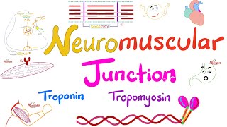
Neuromuscular Junction - Animation

In order for a skeletal muscle to contract, your brain sends a signal, in the form of an action potential in an upper motor neuron.
The upper motor neuron is part of the cerebral cortex, and it activates a lower motor neuron, which is located in the anterior horn of the spinal cord.
From here, the action potential is sent through an axon down to its ending branches, called axon terminals, to muscle fibers which they innervate.
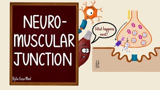
The place where an axon terminal meets the muscle is the neuromuscular junction.
Now, let's take a closer look at neuromuscular junction.
The neuromuscular junction can be divided into three main parts: a presynaptic part which is the membrane of an axon terminal, the postsynaptic part which is the membrane of a skeletal muscle fiber and is also called a motor end plate , and an area between the nerve terminal and motor endplate which is called synaptic cleft.
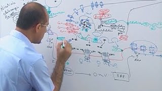
When an action potential spreads over the axon terminal, voltagegated calcium channels open, resulting in an influx of calcium ions into the axon terminal.
Inside the axon terminal are synaptic vesicles that contain neurotransmitters called acetylcholine.
These calcium ions entering the axon terminal bind to vesicles filled with acetylcholine. And this binding allows the vesicles to fuse with the membrane of the axon terminal. And then acetylcholines are released into the synaptic cleft by exocytosis.
The released acetylcholines subsequently binds to nicotinic acetylcholine receptors on the motor endplate. The binding of acetylcholines to receptors triggers the opening of acetylcholine gated ion channels that allow the influx of sodium ions into the muscle.
The principal effect of opening the acetylcholinegated channels is to allow large numbers of sodium ions to pour to the inside of the fiber, carrying with them large numbers of positive charges. The sodium influx changes the postsynaptic membrane potential from negative 90 millivolts to negative 45 millivolts. In turn, this positive potential change inside the muscle fiber membrane initiates an action potential that spreads along the muscle membrane and thus causes muscle contraction.
The acetylcholine, once released into the synaptic cleft, continues to activate the acetylcholine
receptors as long as the acetylcholine persists in the synaptic cleft. To prevent sustained muscle contraction, acetylcholine is removed from the synaptic cleft in 2 ways. Firstly most of the acetylcholine is destroyed by the enzyme acetylcholinesterase which is an enzyme that causes rapid hydrolysis of acetylcholine. And secondly a small amount of acetylcholine diffuses out of the synaptic cleft and is then no longer available to act on the muscle fiber membrane.
With these two ways, acetylcholines in the synaptic cleft are removed. And muscle contraction ends.