Neuromuscular Junction (NMJ) Structure and Action

LIKE US ON FACEBOOK : fb.me/Medsimplified
BUY USING AFFILIATE LINKS :
AMAZON US https://goo.gl/XSJtTx
AMAZON India http://goo.gl/QsUhku
FLIPKART http://fkrt.it/Wiv8RNNNNN'>http://fkrt.it/Wiv8RNNNNN
FLIPKART MOBILE APP http://fkrt.it/Wiv8RNNNNN'>http://fkrt.it/Wiv8RNNNNN
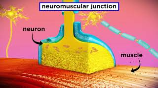
A neuromuscular junction (or myoneural junction) is a chemical synapse formed by the contact between a motor neuron and a muscle fiber.[1] It is at the neuromuscular junction that a motor neuron is able to transmit a signal to the muscle fiber, causing muscle contraction.
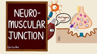
Muscles require innervation to function—and even just to maintain muscle tone, avoiding atrophy. Synaptic transmission at the neuromuscular junction begins when an action potential reaches the presynaptic terminal of a motor neuron, which activates voltagedependent calcium channels to allow calcium ions to enter the neuron. Calcium ions bind to sensor proteins (synaptotagmin) on synaptic vesicles, triggering vesicle fusion with the cell membrane and subsequent neurotransmitter release from the motor neuron into the synaptic cleft. In vertebrates, motor neurons release acetylcholine (ACh), a small molecule neurotransmitter, which diffuses across the synaptic cleft and binds to nicotinic acetylcholine receptors (nAChRs) on the cell membrane of the muscle fiber, also known as the sarcolemma. nAChRs are ionotropic receptors, meaning they serve as ligandgated ion channels. The binding of ACh to the receptor can depolarize the muscle fiber, causing a cascade that eventually results in muscle contraction.
Neuromuscular junction diseases can be of genetic and autoimmune origin. Genetic disorders, such as Duchenne muscular dystrophy, can arise from mutated structural proteins that comprise the neuromuscular junction, whereas autoimmune diseases, such as myasthenia gravis, occur when antibodies are produced against nicotinic acetylcholine receptors on the sarcolemma.
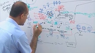
The neuromuscular junction differs from chemical synapses between neurons. Presynaptic motor axons stop 30 nanometers from the sarcolemma, the cell membrane of a muscle cell. This 30nanometer space forms the synaptic cleft through which signalling molecules are released. The sarcolemma has invaginations called postjunctional folds, which increase the surface area of the membrane exposed to the synaptic cleft.[2] These postjunctional folds form what is referred to as the motor endplate, which possess nicotinic acetylcholine receptors (nAChRs) at a density of 10,000 receptors/micrometer2 in skeletal muscle.[3] The presynaptic axons form bulges called terminal boutons (or presynaptic terminals) that project into the postjunctional folds of the sarcolemma. The presynaptic terminals have active zones that contain vesicles, quanta, full of acetylcholine molecules. These vesicles can fuse with the presynaptic membrane and release ACh molecules into the synaptic cleft via exocytosis after depolarization.[2] AChRs are localized opposite the presynaptic terminals by protein scaffolds at the postjunctional folds of the sarcolemma. Dystrophin, a structural protein, connects the sarcomere, sarcolemma, and extracellular matrix components. Rapsyn is another protein that docks AChRs and structural proteins to the cytoskeleton. Also present is the receptor tyrosine kinase protein MuSK, a signaling protein involved in the development of the neuromuscular junction, which is also held in place by rapsyn.[2]
Mechanism of action
The neuromuscular junction is where a neuron activates a muscle to contract. Upon the arrival of an action potential at the presynaptic neuron terminal, voltagedependent calcium channels open and Ca2+ ions flow from the extracellular fluid into the presynaptic neuron's cytosol. This influx of Ca2+ causes neurotransmittercontaining vesicles to dock and fuse to the presynaptic neuron's cell membrane through SNARE proteins. Fusion of the vesicular membrane with the presynaptic cell membrane results in the emptying of the vesicle's contents (acetylcholine) into the synaptic cleft, a process known as exocytosis. Acetylcholine diffuses into the synaptic cleft and can bind to the nicotinic acetylcholine receptors on the motor endplate.
wATCH aGAIN • Neuromuscular Junction (NMJ) Structur...
~~~~~~~~~
CHECK OUT NEWEST VIDEO: "Nucleic acids DNA and RNA structure "
• Nucleic acids DNA and RNA structure
~~~~~~~~~