The easiest way to skyrocket your YouTube subscribers

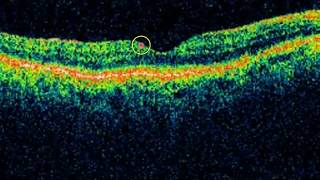
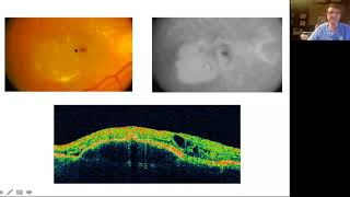
NORMAL MACULAR ANATOMY ON OCT

This video talks about the normal macular and retinal anatomy on OCT scan. the normal macular anatomy on oct can be studied by noticing the vitreoretinal interface, the inner retinal layer, the outer retinal layer and the choroid and the sclera . The VR interface is the junction between the vtreous and the ILM ( internal limiting memberane of the retina. The inner retina consists of layers of hyperreflective and hyporeflective zones on OCT. the Outer retinal layer is again divided into 4 parts ( the outer limiting memberane, ellipsoid zone,interdigitation zone, the RPEBruchs comples. Using the enhanced depth imaging, the choroid and retina can also be images and studies on OCT
#oct
#ophthalmology
#retina


Recommended