Obstetric Ultrasound Normal Vs Subchorionic Hemorrhage (SCH) | Early u0026 Mid Pregnancy Bleeding USG

Obstetric Ultrasound Normal Vs Subchorionic Hemorrhage (SCH) | Early & Mid Pregnancy Bleeding USG
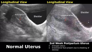
Subchorionic Hemorrhage
A collection of blood between the uterine wall and the gestational sac.
Hypoechoic area between the gestational sac and the uterine wall.
Causes: Trauma, maternal conditions, such as clotting disorders or hypertension, hormonal changes, infections or inflammatory conditions of the reproductive tract, a history of pregnancy complications, such as previous subchorionic hemorrhages or miscarriages
Lack of blood flow on color doppler within the collection can help differentiate it from other types of masses.
Acute SCH appears hyperechoic on ultrasound.
May be difficult to differentiate from the adjacent chorion
SubAcute SCH becomes less echogenic
Chronic SCH appears hypoechoic
Small: Less than 20% of the gestational sac size
Medium: 2050% of the gestational sac size
Large: Greater than 50% of the gestational sac size (Higher risk of complications such as miscarriage & preterm delivery)
Large SCH Complications:
Miscarriage: The risk of miscarriage is a major concern, especially in the first trimester. Studies suggest a higher miscarriage rate associated with larger subchorionic hematomas, particularly when diagnosed early in pregnancy.
Preterm Delivery: Large hematomas can disrupt the vital exchange of nutrients and oxygen between mother and baby. This may increase the risk of preterm labor and delivery, potentially leading to complications for the baby like breathing difficulties or developmental delays
Placental Abruption: In severe cases, a large subchorionic hemorrhage can detach the placenta partially from the uterine wall.