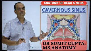
Paired Dural Venous Sinuses-The Cavernous Sinuses |Situation |Relations |Tributaries |Communications

Introduction
Each cavernous sinus is a large venous space situated in the middle cranial fossa,
on either side of the body of the sphenoid bone.
Its interior is divided into a number of spaces or caverns by trabeculae.
The trabeculae are much less conspicuous in the living than in the dead.
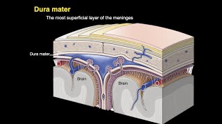
The floor and medial wall of the sinus is formed by the endosteal dura mater.
The lateral wall, and roof are formed by the meningeal dura mater.
Anteriorly, the sinus extends up to the medial end of the superior orbital fissure and
posteriorly, up to the apex of the petrous temporal bone.
It is about 2 cm long, and 1 cm wide.
Relations
Structures outside the sinus:
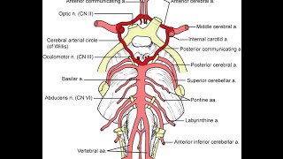
1. Superiorly: Optic tract, optic chiasma, olfactory tract, internal carotid artery and anterior perforated substance.
2. Inferiorly: Foramen lacerum and the junction of the body and greater wing of the sphenoid bone
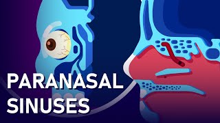
3. Medially; Hypophysis cerebri and sphenoidal air sinus.
4. Laterally: Temporal lobe with uncus.
5. Below laterally: Mandibular nerve
6. Anteriorly; Superior orbital fissure and the apex of the orbit.
7. Posteriorly; Apex of the petrous temporal and the crus cerebri of the midbrain.
Structures with in the Lateral Wall
From above downwards
1. Oculomotor nerve: In the anterior part of the sinus, it divides into superior and inferior divisions which leave the sinus by passing through the superior
orbital fissure.
2. Trochlear nerve: In the anterior part of the sinus, it crosses superficial to the oculomotor nerve, and enters the orbit through the superior orbital fissure.
3. Ophthalmic nerve: In the anterior part of the sinus, it divides into the lacrimal, frontal and nasociliary nerves.
4. Maxillary Nerve: It leaves the sinus by passing through the foramen rotundum on its way to the pterygopalatine fossa.
5. Trigeminal ganglion: The ganglion and its dural cave project into the posterior part of the lateral wall of the sinus. Structures passing through the medial aspect of the sinus: a. lnternal carotid artery with the venous and sympathetic plexus around it.
b. Abducent nerve, inferolateral to the internal carotid artery. The structures in the lateral wall and on the medial aspect of the sinus are separated from blood by the endothelial lining.
Tributaries or Incoming Channels
From the orbit
1. The superior ophthalmic vein.
2. A branch of the inferior ophthalmic vein or sometimes the vein itself.
3. The central vein of the retina may drain either into the superior ophthalmic vein or into the cavernous sinus
From the Brain
1. Superficial middle cerebral vein.
2. Inferior cerebral veins from the temporal lobe From the Meninges
1. Sphenoparietal sinus.
2. The frontal trunk of the middle meningeal vein may drain either into the pterygoid plexus through the foramen ovale or into the sphenoparietal or cavernous sinus.
Draining Channels or Communications
The cavernous sinus drains:
1. Into the transverse sinus through the superior petrosal sinus.
2. Into the internal jugular vein through the inferior petrosal sinus and through a plexus around the internal carotid artery.
3. Into the pterygoid plexus of veins through the emissary veins passing through the foramen ovale, the foramen lacerum and the emissary sphenoidal foramen.
4. Into the facial vein through the superior ophthalmic vein.
5. The right and left cavernous sinuses communicate with each other through the anterior and posterior intercavernous sinuses and through the basilar plexus of veins
All these communications are valveless, and blood can flow through them in either direction.
Factors helping expulsion of blood from the Sinus
1. Expansile pulsations of the internal carotid artery within the sinus. 2. Gravity.
3. Position of the head.
Clinical Anatomy
1. Thrombosis of the cavernous sinus may be caused by sepsis in the dangerous area of the face, in nasal cavities, and in paranasal air sinuses. This gives rise to the following symptoms.
a. Nervous symptoms:
Severe pain in the eye and forehead in the area of distribution of ophthalmic nerve.
Involvement of the third, fourth and sixth cranial nerves resulting in paralysis of the muscles supplied.
b. Venous symptoms: Marked oedema of eyelids, cornea and root of the nose, with exophthalmos due to congestion of the orbital veins.
2. A communication between the cavernous sinus and the internal carotid artery may be produced
by head injury. When this happens the eyeball protrudes and pulsates with each heart beat. It is called the pulsating exophthalmos.
Follow me in blogspot https://humananatomylessons.blogspot... _________________________________________________________________________________________________________________ Visit my blogs https://humananatomyonline.in/
Contact me @ https://t.me/humananatomylessons