Parietal Bone - Definiton Location u0026 Sutures - Human Anatomy | Kenhub

This video covers the anatomy of the parietal bone: parietal bone location, landmarks, sutures and articulations. Take our quiz on the lateral and posterior view of the skull at https://khub.me/o2bef
Oh, are you struggling with learning anatomy? We created the ★ Ultimate Anatomy Study Guide ★ to help you kick some gluteus maximus in any topic. Completely free. Download yours today: https://khub.me/bnvug
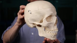
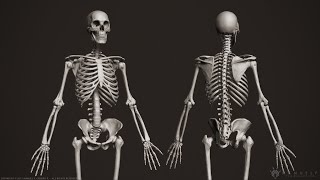
The parietal bone is a roughly squareshaped, paired bone that forms large parts of the top and side of the head.
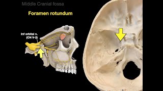
Its concave internal surface is covered with several grooves and landmarks for other structures inside the skull, such as the middle meningeal artery, the sigmoid sinus and the superior sagittal sinus. The convex external surface shows a parietal foramen, through which a parietal emissary vein connects the veins of the scalp with the superior sagittal sinus.
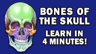
The two parietal bones meet each other in the midline of the skull roof, forming a serrated margin known as the sagittal suture. Apart from its opposite counterpart, each parietal bone is surrounded by 4 other bones: frontal bone, occipital bone, temporal bone and the sphenoid. It is connected to each bone by a suture.
In this tutorial we will go over the most relevant facts about the parietal bone anatomy:
0:09 parietal bone location and shape
0:33 internal and external surface of the parietal bone
1:13 parietal bone sutures
Want to test your knowledge on the structures seen on the anterior view of the skull? Take this quiz: https://khub.me/nheyc
Read more on the components, foramina, cranial fossae and bones of the skull in our free article and step up your skull game: https://khub.me/lw69l
For more engaging video tutorials, interactive quizzes, articles and an atlas of Human anatomy and histology, go to https://khub.me/2zh2n


![Skull Bone & Suture Mnemonic/Trick [Cranial Bone Anatomy Animation]](https://i.ytimg.com/vi/P21JpzKtlxE/mqdefault.jpg)


![Facial Bones of the Skull Mnemonic [Anatomy Animation]](https://i.ytimg.com/vi/d2D-OMUD2q0/mqdefault.jpg)