Parotid Gland | Parotid mould or Bed | Parts | Relations | Blood supply | Secretomotor Pathway

The parotid is the largest of the salivary glands.
• It weighs about 15 g.
• It is situated below the external acoustic meatus, between the ramus of the mandible and the
sternocleidomastoid. The gland overlaps these structures.
• Anteriorly, the gland also overlaps the masseter muscle.
• A part of this forward extension is often detached, and is known as the accessory parotid, and it lies between the zygomatic arch and the parotid duct.
Capsule of Parotid Gland
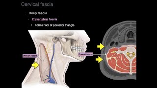
• The investing layer of the deep cervical fascia forms a capsule for the gland.
• The fascia splits (between the angle of the mandible and the mastoid process) to enclose the gland.
• The superficial lamina, thick and adherent to the gland, is attached above to the zygomatic arch.
• The deep lamina is thin and is attached to the styloid process, the angle and posterior border of the ramus of the mandible and the tympanic plate.
• A portion of the deep lamina, extending between the styloid process and the mandible, is thickened to form the stylomandibular ligament which separates the parotid gland from the submandibular salivary gland.
• The ligament is pierced by the external carotid artery
Clinical Anatomy
• Parotid swellings are very painful due to the unyielding nature of the parotid fascia.
• Mumps is an infectious disease of the salivary glands (usually the parotid) caused by a specific virus,
Viral parotitis or mumps characteristically does not suppurate. Its complications are orchitis and pancreatitis
External Features
The gland resembles a three sided pyramid.
The apex of the pyramid is directed downwards
The gland has four surfaces:
a. Superior (base of the pyramid)
b. Superficial
c. Anteromedial
d. Posteromedial.
The surfaces are separated by three borders:
a. Anterior
b. Posterior
c. Medial/pharyngeal
Relations
The apex overlaps the posterior belly of the digastric and the adjoining part of the carotid triangle.
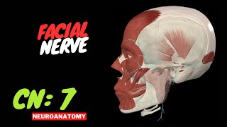
The cervical branch of the facial nerve and the two divisions of the retromandibular vein emerge near the apex.
Surfaces
The superior surface or base forms the upper end of the gland which is small and
concave.
It is related to:
a. The cartilaginous part of the external acoustic meatus.
b. The posterior surface of the temporomandibular joint.
c. The superficial temporal vessels.
d. The auriculotemporal nerve
The superficial surface is the largest of the four surfaces.
It is covered with:
a. Skin
b. Superficial fascia containing the anterior branches of the great auricular nerve, the
preauricular or superficial parotid lymph nodes and the posterior fibres of the
platysma and risorius.
c. The parotid fascia which is thick and adherent to the gland.
d. A few deep parotid lymph nodes embedded in the gland
The anteromedial surface is grooved by the
posterior border of the ramus of the mandible.
It is related to:
a. The masseter
b. The lateral surface of the temporomandibular joint.
c. The posterior border of the ramus of the mandible
d. The medial pterygoid
e. The emerging branches of the facial nerve.
The posteromedial surface is moulded to the mastoid and the styloid processes and the
structures attached to them.
Thus it is related to:
a. The mastoid process, with the sternocleidomastoid and the posterior belly of the digastric.
b. The styloid process, with structures attached to it.
c. The external carotid artery enters the gland through this surface and the internal carotid artery lies deep to the styloid process
Borders
The anterior border separates the superficial surface from the anteromedial surface.
It extends from the anterior part of the superior surface to the apex.
The following structures emerge at this border:
a. The parotid duct.
b. Most of the terminal branches of the facial nerve.
c. The transverse facial vessels. In addition, the accessory parotid gland lies on the parotid duct
close to this border.
The posterior border separates the superficial surface from the posteromedial surface. It overlaps the sternocleidomastoid.
The medial edge or pharyngeal border separates the anteromedial surface from the posteromedial surface. It is related to the lateral wall of the pharynx
Structures within the parotid gland
From medial to the lateral side, these are as follows.
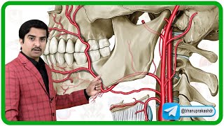
Arteries: The external carotid artery enters the gland through its posteromedial surface.
The maxillary artery
The superficial temporal artery
Veins: The retromandibular vein
My website https://humananatomylessons.busines...
My Blog https://humananatomylessons.blogspot...
Follow me in blogspot https://humananatomylessons.blogspot...