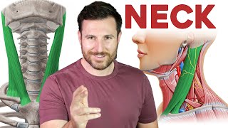
Posterior triangle of the neck

: / drgbhanuprakash
POSTERIOR TRIANGLE OF NECK
The posterior triangle of the neck forms the posterior compartment of the neck and is separated from the anterior triangle by the sternocleidomastoid muscle. The triangles of the neck are surgically focused, first described from early dissectionbased anatomical studies which predated crosssectional anatomical description based on imaging (see deep spaces of the neck).
Boundaries
anterior: posterior border of sternocleidomastoid
posterior: anterior border of trapezius
inferior: middle third of the clavicle
roof: skin, superficial fascia and the investing layer of deep cervical fascia
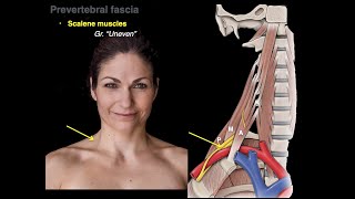
floor: prevertebral fascia overlying splenius capitis, semispinalis capitis, levator scapulae, scalenus medius and scalenus anterior
The inferior belly of the omohyoid that crosses the triangle divides it into an inferior supraclavicular and superior occipital triangles.
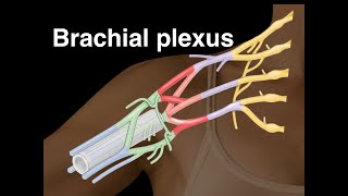
Contents
The posterior triangle contains level 5 lymph node chains. These include spinal accessory and transverse cervical nodes. Depending on the location of the nodes above or below the accessory nerve, they are sub grouped as level 5a (above) or level 5b (below).
#usmle #usmlestep1 #nationalexittest #usmlevideos #usmlepreparation #usmleprep #usmlecoaching #neetpg #neetpgvideos #marrow #prepladder #drbhanuprakash #anatomy #anatomylecture #anatomyvideos