Red Blood Cell Morphological Abnormalities

This is a video describing various morphological abnormalities found in red blood cells, including changes in red cell size, shape, color, and distribution.
I created this presentation with Google Slides.
Image were created or taken from Wikimedia Commons
I created this video with the YouTube Video Editor.
ADDITIONAL TAGS:
Macrocytosis
Large
As measured with MCV
altered DNA synthesis
MCV,
things that alter DNA synthesis , B12/folate deficiency
Liver disease
Thyroid disease
Chemotherapy
Antiretrovirals (AZT)
Microcytosis
Small
Measured with MCV
iron deficiency, thalassemia, lead poisoning
measured with MCV,
Iron deficiency, Thalassemias, Lead poisoning, Sideroblastic anemia
Anisocytosis
Wide range of RBC sizes
High red cell distribution width (RDW)
Hypochromasia
with too little hemoglobin
Measured with mean corpuscular Hb (MCH)
hypochromic cells, central cell diameter
caused by lack of hemoglob (iron defiiciency, thalassemia, liver problems)
Polychromasia
that are shaded grayish blue
often reticulocytes (immature )
Anisocytosis, large RDW
Poikilocytosis
that vary widely shape
Analogous anisocytosis
anisocytosis vary size
poikilocytosis vary shape
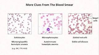
Target cells
codocytes
Related liver disease, thalassemias, hemoglob C, postsplenectomy
Normal RBC
cross section
Target cell
Anisocytosis, large RDW
Spherocytes
Spherical (instead of biconcave diskshaped
Appears on blood smear as loss of central pallor
Hereditary spherocytosis
Autoimmune hemolysis
Normal RBC Spherocyte
cross section cross section
(biconcave disk) (more spherical)
Ab attack membrane of RBC, if you pull out chunks of membrane, surface area of RBC membrane decreases
Schistocytes
fragments
Sharp edges
Can be caused by plaque on arterial walls that shear
Also Microangiopathic Hemolytic Anemia (MAHA)
Sickle cells
fragments
sickle cell anemia
Hemoglob molecules with
Point mutation that changes glutamic acid valine
Polymerization of Hb at low pH, low pO2, high temperature
Anisocytosis, large RDW
Echinocytes
burr cells
Projections that are regular
renal disease
Acanthocytes
spur cells
Projections that are irregular large
liver disease
Teardrop cells
dacrocytes
Caused by infiltration of bone marrow (myelophthisic processes)
Something body ( lymphocytes, scar tissue, cancer) enters grows bone marrow
Linear aggregations or “stacks” of
are high levels of immunoglobulins
usually have similar surfaces charges that keep them from sticking together
Immunoglobulins neutralize these charges, allow attract with chargecharge interactions
a normal patient, you might see this on dense side of a blood smear, bc RBC conc is more concentrated
Agglutination
collect clumps
Less orderly than rouleaux
are coated with IgM, which bridge together cause aggregation
HowellJolly Bodies
Remnants of nucleic acids that found postsplenectomy
Look like little purple dots