Sacral Plexus Anatomy Animation : Dr G Bhanu Prakash

: / drgbhanuprakash
: https://t.me/bhanuprakashdr
: https://linktr.ee/DrGBhanuprakash
Sacral Plexus Anatomy Animation : Dr G Bhanu Prakash
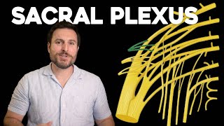
Sacral Plexus Anatomy
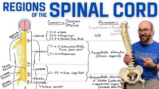
The Sacral Plexus is anatomically located within the pelvis, formed by the converging anterior rami of the L4 to S4 spinal nerves. This plexus is positioned on the pelvic posterior wall, lying anterior to the piriformis muscle. The nerve roots from the lumbar and sacral levels intricately interconnect to create a network that gives rise to several crucial nerves. Key branches include the sciatic nerve, which is the largest and is responsible for innervating the hamstrings, adductor magnus, and the majority of the leg and foot muscles. It also provides sensory innervation to the skin of the foot and lower leg. The pudendal nerve caters to the perineal muscles, external genitalia, and the skin around the anus and perineum, playing a significant role in the mechanisms of urination and defecation. The superior and inferior gluteal nerves innervate the gluteal muscles, with the former activating the gluteus medius, gluteus minimus, and tensor fasciae latae, while the latter is dedicated to the gluteus maximus.
In terms of its relationships with surrounding structures, the sacral plexus is intimately associated with the pelvic organs and is located near the sacroiliac joint. The sciatic nerve notably exits the pelvis through the greater sciatic foramen below the piriformis muscle. Clinically, the relevance of the sacral plexus is highlighted in conditions such as sciatica, where pain is experienced along the sciatic nerve's distribution, often due to a herniated disc, and pudendal neuralgia, which involves chronic pelvic pain related to the pudendal nerve.
The plexus is vascularized by branches of the internal iliac artery and has venous and lymphatic drainage systems that follow the arterial layout, draining into the internal iliac and sacral lymph nodes. From an embryological standpoint, the sacral plexus forms alongside the development of the lower limb buds, originating from the ventral divisions of the spinal nerves. It is not uncommon to encounter anatomical variations in the sacral plexus, which can influence the clinical manifestations of nerverelated injuries.
Diagnostic evaluation of the sacral plexus typically involves MRI for detailed imaging, while electrophysiology through electromyography may be utilized to assess muscular function attributed to the sacral plexus innervation. Surgical interventions in the pelvis necessitate a comprehensive understanding of sacral plexus anatomy to prevent iatrogenic nerve damage, which is paramount to preserving lower limb and pelvic functions. Rehabilitation often includes physical therapy to strengthen affected muscles and various pain management techniques for neuropathic conditions arising from disorders of the plexus.
#fmge #fmgevideos #rapidrevisionfmge #fmgejan2023 #mbbslectures #nationalexitexam #nationalexittest #neetpg #usmlepreparation #usmlestep1 #fmge #usmle #drgbhanuprakash #medicalstudents #medicalstudent #medicalcollege #neetpg2023 #usmleprep #usmlevideos #usmlestep1videos #medicalstudents #neetpgvideos #sacralplexus #drgbhanuprakash #drbhanuprakash