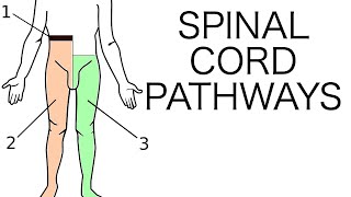
Spinal Cord: Anatomy Spinal Tracts u0026 Pathways Somatic Reflexes Animation

(USMLE topics, brain and nervous system)
Purchase a license to download a nonwatermarked version of this video on AlilaMedicalMedia(dot)com
Check out our new Alila Academy AlilaAcademy(dot)com complete video courses with quizzes, PDFs, and downloadable images.
©Alila Medical Media. All rights reserved.
Voice by: Ashley Fleming
All images/videos by Alila Medical Media are for information purposes ONLY and are NOT intended to replace professional medical advice, diagnosis or treatment. Always seek the advice of a qualified healthcare provider with any questions you may have regarding a medical condition.
The spinal cord is a long, thin tube of nervous tissue, enclosed in 3 membranes of the meninges which, in turn, are protected within the bones of the vertebral column. The 31 pairs of spinal nerves arise from the cord and emerge from the vertebrae. The spinal cord extends from the brainstem to the level of upper lumbar vertebrae. In the lower lumbar and sacral regions, nerve roots descend within the spinal canal before exiting, forming the cauda equina.
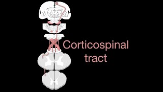
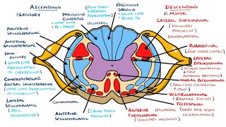
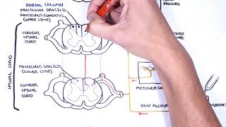
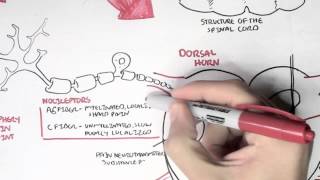
In cross section, two types of nervous tissue can be seen in the cord: a butterflyshaped central core of gray matter, and a surrounding white matter. The gray matter contains cell bodies and dendrites of neurons. This is where neurons synapse and transmit information to each other. The white matter, on the other hand, is made of bundles of axons, and serves to conduct information up and down the cord. These bundles are organized into specific groups with specific functions, forming the socalled spinal tracts. Ascending tracts conduct sensory information up to the brain, while descending tracts convey motor instructions down the cord. Some tracts cross over to the other side of the cord, before they reach the brain. They convey sensory information from one side of the body to the other side of the brain. When this happens, the information is said to be transmitted contralaterally. Tracts that stay on the same side all the way are said to conduct information ipsilaterally.
A sensory pathway typically involves 3 neurons:
Firstorder neurons detect stimuli and transmit signals to the spinal cord. The axons of these neurons form sensory fibers that enter the cord via the dorsal root of spinal nerve.
Inside the cord, firstorder neurons synapse with secondorder neurons, which ascend a specific tract to the brainstem, or further up to the thalamus. In some pathways, firstorder neurons ascend the tract to the brainstem, where they synapse with secondorder neurons, which continue to the thalamus.
Thirdorder neurons conduct the information the rest of the way to the sensory cortex.
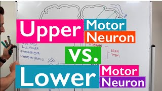
A motor pathway usually involves 2 neurons: an upper motor neuron starts in the motor cortex or brainstem, and a lower motor neuron continues from the brainstem or spinal cord. They conduct motor instructions down, along a specific descending tract. The axons of lower motor neurons exit the cord via the ventral root of spinal nerve, where they continue as motor fibers to effector organs.
The spinal cord is also responsible for fast, involuntary responses of skeletal muscles, called somatic reflexes. Reflexes are essentially automatic and do not require input from the brain, although the brain is informed and aware, usually afterthefact. A somatic reflex involves a reflex arc composed of a somatic receptor, a sensory neuron, an interneuron, a motor neuron, and an effector muscle. Some reflexes are however more complex, and require multiple pathways, as well as central coordination from the brain.