Structure and Function of EYES (in Hindi)

In this Biology video in Hindi we explained the basic structure and function of human eye. We discussed on the different parts of eye and their individual functions.
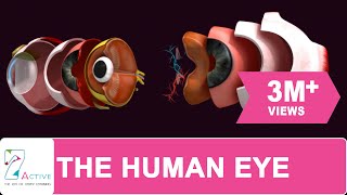
Human eye has many parts for different functions. Following are the most important parts of eye.
1. Sclera : It is the outer protective layer of eye
2. Iris : It is the colored part of eye, visible from outside.
3. Pupil : It is the central black part inside the iris.
4. Cornea : It is the outer protective layer of iris.
5. Anterior chamber : It is the chamber between iris and cornea. It is filled with a fluid called aqueous humour.
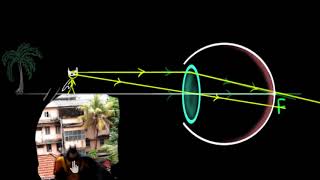
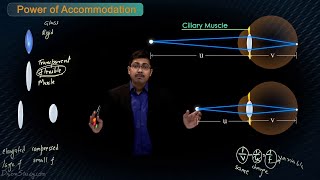
6. Lens : It is situated behind the iris to converge the light rays onto the retina to create the image of an object. It can be contracted or elongated with the help of a muscle named ciliary body.
7. Exterior chamber : It is the chamber between iris and the lens. It is filled with a fluid called aqueous humour.
8. Vitreous humour : It is a fluid situated between the lens and the retina.
9. Choroid : It is a layer inside the sclera.
10. Retina : It is a layer containing the photoreceptor cells.
11. Cone cells : These photoreceptor cells can detect colour in bright light. When tens and hundreds of photons incident on a cone cell, it activates.
It is of three types, viz.,
First, having blue sensitive opsin protein named OPN1SW, which can be triggered if blue light incidents on it.
Second, having green sensitive opsin protein named OPN1MW, which can be triggered if green light incidents on it.
Third, having red sensitive opsin protein named OPN1LW, which can be triggered if red light incidents on it.
12. Rod cells : These photoreceptor cells can detect low light without the sensation of colour. When only one photon incidents on a rod cell, it activates. Hence, it is responsible for low light vision, esp., at night.
It contains a protein called Rhodopsin, which contains a derivative of Vitamin A. That is why the deficiency of Vitamin a leads to night blindness.
13. Fovea : It is the part of the retina just behind the lens. It contains only cone cells in very high density. The density of cone cell reduces as we go further the fovea. That is why we can see an object just in front of our eyes very clearly. But as we go further away the visual clarity fades away.
14. Blind spot : The place where optic nerve and retinal blood vessels enter the eye, there are no photoreceptor cells there. Hence, when light incidents on that region, no visual sensation is created.
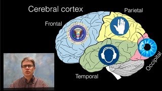
15 : Optic nerve : When photoreceptor cells create impulse, that impulse is carried by these nerves to the occipital lobe of the brain, where the sense of vision is produced.
16. Retinal blood vessels : These blood vessels carry blood to the retina.