Tarsal Coalition - Everything You Need To Know - Dr. Nabil Ebraheim

Dr. Ebraheim’s educational animated video describes the accessory navicular bone.
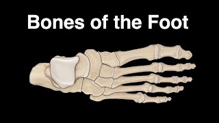
Tarsal coalition is fusion of the tarsal bones that leads to a rigid flat foot, foot pain and multiple ankle sprains. It is a congenital anomaly.
Two types of tarsal coalition:
1Talocalcaneal Coalition: it is coalition between the talus and the calcaneus. When talocalcaneal coalition occurs, it usuaully happens around 1215 years of age.
2Calcaneonavicular coalition: it is a coalition between the calcaneus and the navicular. The calcaneonavicular coalition presents at an earlier age.
50% of coalitions are bilateral. About 20% have multiple coalition in the same foot. Coalition may be fibrous, cartilaginous or bony. It occurs due to failure of segmentation. It could be associated with fibular hemimelia or Apert’s syndrome.
Symptoms:
•the patient usually complains of a painful foot.
•A history of repeated ankle sprains.
•Flatfoot deformity
Examination: Tarsal coalition may result in peroneal spastic flat foot. You may also find hindfoot valgus. On toe standing, the arch does not reconstitute. Heel cord contracture. Restricted subtalar joint range of motion. Always check both feet, the condition may be bilateral.
The best study is CT scan. It can determine the size and location of coalition. MRI is also useful in detecting a fibrous or cartilaginous coalition. Xrays: AP view, lateral view and oblique view.
Lateral view xrays:
•Calcaneonavicular coalition “anteater nose sign” is elongation of the anterior calcaneal process.
•Talocalcaneal coalition: talar breaking may be shown on lateral radiographs. It is traction spur that occurs due to limited motion of the subtalar joint. The C sign is a radiological sign which may be seen on lateral radiographs. It is the outline of the talar dome and sustentaculum.
Oblique view xrays: 45 degree oblique view is the best for showing calcaneonavicular coalition.
Treatment:
•NSIADS
•MODIFIED ACTIVITIES
•BRACE OR CAST.
•Surgery: resection of the calcaneonavicular coalition with interposition of the extensor digitorum brevis muscle or a fat graft no matter what the size of coalition is. Talocalcaneal coalitions that involve less than 50% of the subtalar joint are also resected. A triple arthrodesis is performed for large coalitions, failed resections, or advanced conditions.
Become a friend on facebook:
/ drebraheim
Follow me on twitter:
https://twitter.com/#!/DrEbraheim_UTMC
Donate to the University of Toledo Foundation Department of Orthopaedic Surgery Endowed Chair Fund:
https://www.utfoundation.org/foundati...


![Accessory Navicular Bone Pain [Inside of Foot Pain, Inside of Arch]](https://i.ytimg.com/vi/HnHY9Cgn7jU/mqdefault.jpg)