The Anatomy of Thoracic Outlet Syndrome: The Anterior Scalene Muscle is the Key to TOS

Join Dr. Scott Werden for the most recent in his "The Anatomy of Thoracic Outlet Syndrome" series as he discusses The Anterior Scalene Muscle and why it is the key to understanding TOS. Live participant Q & A.
https://www.toseducation.org/videoop...
https://www.tosmri.com/thoracicoutle...
thoracic outlet syndrome symptoms
thoracic outlet syndrome test
thoracic outlet syndrome
thoracic outlet syndrome exercises
thoracic outlet syndrome specialist
don't guess with tos
the tos guy
do I have thoracic outlet syndrome
do I have TOS
thoracic outlet syndrome education
Understanding anatomy is the key to understanding TOS
Abnormal anatomy causes or contributes to compression of the vital structures in the thoracic outlet. As a result, patients experience the symptoms of TOS. While the anatomy of the thoracic outlet is complex, understanding the anatomy enables understanding of TOS. We can strive to understand the structures that cause compression as well as the structures that become compressed.
We can categorize the essential anatomy into these categories:
Anatomy of the spine
Anatomy of a nerve
Anatomy of the brachial plexus
Anatomy of the thoracic outlet
Anatomy of the spine
The spine supports our body weight while allowing flexibility in multiple directions.
The vertebral body is the basic building block of the spine. Vertebral bodies (also known as vertebrae) are stacked one upon the other from the skull to the pelvis. The vertebral bodies support our body weight.
Between each vertebral body lies an intervertebral disc. Each disc is firmly attached to the vertebral body above it and to the body below it. The disc has a fibrous outer layer that stabilizes each vertebral body to the next. The center of each disc contains a jellylike substance that absorbs impacts from walking, jumping and other movement. The flexible discs allow movement in multiple directions:
Flexion and extension: Forward and backward bending
Lateral bending: Side to side bending
Rotation: Side to side twisting
Vertebral bodies and intervertebral discs
The spine comprises five anatomic regions, from top to bottom:
Cervical spine: The neck
Thoracic spine: The midback; twelve paired ribs arise from the thoracic spine, forming the rib cage
Lumbar spine: The low back
Sacral spine: The base of the spine; joins the pelvis to the spine
Coccyx: The tailbone; in humans, the coccyx represents a vestigial tail
Spine anatomy regions
Each region of the spine has a different number of vertebral bodies:
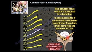
Cervical spineseven segments, numbered C1 (‘Cervical’ 1) through C7
Thoracic spinetwelve segments, numbered T1 (‘Thoracic’ 1) through T12; each body has a rib on each side
Lumbar spinefive segments, numbered L1 (‘Lumbar’ 1) through L5
Sacral spinefive segments, numbered S1 (‘Sacral’ 1) through S5; these segments are fused together into one bone in adults, which articulates with the pelvis on each side.
Coccyxthree to five segments, with variable degrees of fusion in the adult; usually referred to as a single unit rather than separate vertebrae.
A bony arch arises from the back of each vertebral body. These arches line up to form a flexible bony tunnel, the spinal canal. The spinal canal runs from the base of the skull to the pelvis, and it contains and protects the spinal cord. At each spinal level, a nerve leaves the spinal cord on each side. Each nerve exits the spinal canal through a tunnel between the bony arches to reach the body or extremities. This tunnel is called a neural foramen.
On the back and sides of each arch, bony protrusions arise, which allow for attachments of the numerous muscles that stabilize and move the spine.


![TherEx | Scalene Stretches [for Anterior, Middle, & Posterior Scalenes]](https://i.ytimg.com/vi/4WUiHGuENFU/mqdefault.jpg)