This Video Will SAVE 20 HOURS of Your Reading Time || Hemostasis: Animation Series Compilation

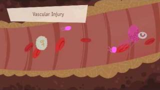
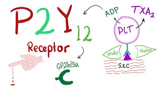
In this video entire physiology of hemostasis starting from platelet plug formation to coagulation cascade to clot retraction and fibrinolysis / clot dissolution is covered with animation. There are three main steps in platelet plug formation. Platelet adhesion, which is initial covering of wound area by platelets. It starts with release of von Willebrand factor by damaged endothelial cells. They bind to the collagen that is exposed as result of vessel injury. Platelet adhesion is mediated by platelet receptor glycoprotein Ib (Gp Ib) which bind with Von Willebrand factor and glycoprotein Ia/IIa and glycoprotein VI both of which bind with collagen. This binding activates platelet and it releases ADP, fibrinogen and some other important factors from their α and dense granules. They freshly synthesize and release thromboxaneA2. Also thrombin is generated on platelet surface due to simultaneous activation of coagulation cascade. Platelet changes shape to increase the surface area for binding with more platelets. Finally, glycoprotein IIb/IIIa undergo conformational change and bind with fibrinogen. This is followed by next step, platelet aggregation. ADP, TxA2 and thrombin activate more circulating platelets by acting on P2Y12, TP and PAR1 respectively. Platelets, activated thus, release more ADP, TxA2 and thrombin to activate even more platelets and meanwhile also bind with other activated platelets with help of Gp IIb/IIIa/. Here fibrinogen and Von Willebrand factor serve as bridge to connect platelets. In this way platelets are recruited till the break in vessel is plugged. This initial plug is called primary platelet plug or primary haemostatic plug. It is stabilized with help of fibrin that is generated at the end of parallelly occurring coagulation cascade. Coagulation usually occurs parallel to the platelet plug formation. It lay lays down fibrin mesh for clot formation. It starts by two pathway. Intrinsic and extrinsic. Intrinsic pathway starts when blood comes in contact with negatively charged surface. Extrinsic pathway starts when tissue factor is exposed to the blood. Both the pathways merge at common pathway i.e. synthesis of factor Xa. It activates prothrombin into thrombin which converts fibrinogen into fibrin monomers. Fibrin monomers polymerize spontaneously and form fibrin strands. Factor XIIIa cause crosslinking between fibrin polymers and forms mesh. Thus we have a stable clot. During the process other blood cells like red blood cell and white blood cells are also trapped in the clot. Arterial clots are rich in platelets and venous clots are rich in fibrin. Clot retraction occurs by interaction of actin and myosin filaments in platelets. During the process clot oozes serum. Serum is basically plasma without clotting factors. It lacks clotting factors as they are used up in clot formation. So, it cannot coagulate. Eventually the break in vessel is repaired, so clot starts to dissolve. The process starts with tissue plasminogen activator(tPA) which activates plasminogen that was trapped in clot when clot was being formed. Thus, the inactive plasminogen is converted to active plasmin. Plasmin degrades fibrin meshwork into fibrin degradation products. This leads to dissolution of clot. Fragments of clot are cleared by phagocytes. Plasmin also prevents unnecessary clot formation as healthy sites. Its nonspecific and can also degrade other clotting factors. So, its activity at healthy sites is kept under control by plasminogen activator inhibitor which inhibit plasminogen activator and α2 antiplasmin which inhibits plasmin. At clot, fibrin potentiates activity of plasminogen activator as well as prevents active plasmin from inhibitory effect of α2antiplasmin by occupying its active site. So, at clot, plasmin activity is higher.
Chapters
00:00 Introduction
00:17 Platelet plug formation
04:59 Coagulation
08:54 Clot dissolution
13:13 Summary