Ulnar Nerve Clinical Examination - Everything You Need To Know - Dr. Nabil Ebraheim

Dr. Ebraheim’s educational animated video describes the Anatomy and the pathway of the Ulnar Nerve.
There are several clinical examinations that are commonly used to test the function and integrity of the ulnar nerve.
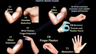
The first muscle that is innervated by the ulnar nerve is the flexor carpi ulnaris.
1 Test flexion of the wrist against resistance: to test the function of the flexor carpi ulnaris muscle, have the patient flex the wrist against resistance in an ulnar direction.
2 Test flexion of the 5th finger against resistance: to test the function of the flexor digitorum profundus, have the patient flex the DIP joint of the 5th finger against resistance, the medial part of the flexor digitorum profundus is supplied by the ulnar nerve.
3 Test abduction of the little finger against resistance: the abductor digiti minimi allows for abduction of the little finger away from the other fingers, the function of the abductor digiti minimi is tested by asking the patient to abduct the little finger against resistance.
4 Test abduction of the index finger against resistance: the function of the dorsal interossei is tested by asking the patient to abduct the index finger against resistance, the first dorsal interossei muscle can be seen and evaluated on the dorsum of the hand, severe atrophy of the first dorsal interosseous muscle could indicate a bad prognisis for recovery of the ulnar nerve, the condition could be associated with a claw hand deformity.
5 Check for clawing of the 4th and 5th fingers: the ulnar claw hand deformity is a symptom of lower ulnar nerve entrapment (below the elbow) and typically causes flexion and clawing of the 4th and 5th fingers due to the unopposed action of the medial part of the flexor digitorum profundus muscle.
6 Froment’s sign: when the adductor pollicis muscle is weak, thumb adduction will not occur, the froment’s sign is used to test the function of the adductor pollicis muscle, when pinching a piece of paper between the thumb and the index finger, the thumb IP joint will flex if the adductor pollicis muscle is weak .
7 Unable to cross the middle and index fingers: as a result of ulnar nerve entrapment and injury the patient is unable to cross or abduct the fingers, adduction of the fingers come from the palmar (PAD) Interossei, abduction of the fingers come from the dorsal (DAB) Interossei.
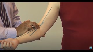
At the elbow, the ulnar nerve travels through a tunnel of tissue (cubital tunnel) that runs under the medial epicondyle.
Pressure on the nerve at the elbow can cause numbness or pain in the elbow, hand, wrist or fingers.
Tinnel’s sign: a tapping technique performed to test for symptoms of ulnar nerve entrapment at the elbow(cubital tunnel syndrome).
Elbow flection test: the elbow flexion test os used to check for symptoms of cubital tunnel syndrome, the patient is asked to fully flex the elbow with the shoulder in some abduction, as the elbow flexes, the area of the cubital tunnel becomes narrow and compresses the nerve, also holding this position as shown, may result in tingling or paresthesia in the ulnar nrve distribution of the forearm or hand, in addition to elbow flexion, adding wrist flexion in an ulnar direction will aggravate the symptoms of cubital tunnel syndrome and induce paresthesia due to contraction of the flexor carpi ulnaris muscle.
Ulnar nerve also provides sensory innervation parts of the hand as distributes in the video.
Become a friend on facebook:
/ drebraheim
Follow me on twitter:
https://twitter.com/#!/DrEbraheim_UTMC
![Hand of Benediction vs the Claw Hand [feat. the Ulnar Paradox]](https://i.ytimg.com/vi/BWhB_B3e4gc/mqdefault.jpg)


![Branches of the Ulnar & Median Nerves [in Hand]](https://i.ytimg.com/vi/ElRwFzT_I58/mqdefault.jpg)