Unpaired Dural Venous Sinuses | Situation | Tributaries | Communications | Confluence of sinuses |

Superior Sagittal Sinus
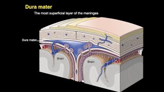
The superior sagittal sinus occupies the upper convex, attached margin of the falx cerebri
It begins anteriorly at the crista galli by the union of tiny meningeal veins.
Here it communicates with the veins of the frontal sinus, and occasionally with the veins of the nose, through the foramen caecum.
As the sinus runs upwards and backwards, it becomes progressively larger in size.
It is triangular on cross section.
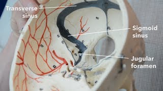
It ends near the internal occipital protuberance by turning to one side, usually the right, and becomes
continuous with the right transverse sinus
It generally communicates with the opposite sinus.
The junction of all these sinuses is called the
confluence of sinuses.
The interior of the sinus shows:
a. Openings of the superior cerebral veins.
b. Openings of venous lacunae, usually three on each side.
c. Arachnoid villi and granulations projecting into the lacunae as well as into the sinus d. Numerous fibrous bands crossing the inferior angle of the sinus.
Tributaries
The superior sagittal sinus receives these tributaries.
a. Superior cerebral veins which never open into the venous lacunae.
b. Parietal emissary veins.
c. Venous lacunae, usually three on each side which first, receive the diploic and meningeal veins, and then open into the sinus.
d. Occasionally, a vein from the nose opens into the sinus when the foramen caecum is patent. Clinical Anatomy:
1. Thrombosis of the superior sagittal sinus maybe caused by spread of infection from the nose, scalp and
diploe.
This gives rise to:
a. A considerable rise in intracranial tension due to defective absorption of CSF.
b. Delirium and sometimes convulsions due to congestion of the superior cerebral veins.
c. Paraplegia of the upper motor neuron type due to bilateral involvement of the paracentral lobules of cerebrum where the lower limbs and perineum are represented.
Inferior Sagittal Sinus
The inferior sagittal sinus, a small channel
lies in the posterior twothirds of the lower, concave free margin of the falx cerebri.
It ends by joining the great cerebral vein to form the straight sinus
Straight Sinus
The straight sinus lies in the median plane within the junction of falx cerebri and the tentorium cerebelli.
It is formed anteriorly by the union of the inferior sagittal sinus with the great cerebral vein, and ends at the internal occipital protuberance by continuing as the transverse sinus usually left In addition to the veins forming it, it also receives a few of the superior cerebellar veins.
At the termination of the great cerebral vein into the sinus, there exists a ball valve mechanism, formed by a sinusoidal plexus of blood vessels, which regulates the secretion of CSF.
Other Sinuses
1. The occipital sinus is small, and lies in the attached margin of the falx cerebelli.
• It begins near the foramen magnum and ends in the confluence of
sinuses
2. The sphenoparietal sinuses, right and left lie along the posterior free margin of the lesser wing of the sphenoid bone, and drain into the anterior part of the cavernous sinus.
• Each sinus may receive the frontal trunk of the middle meningeal vein
3. The superior petrosal sinuses lie in the anterior part of the attached margin of the tentorium cerebelli along the upper border of the petrous temporal bone.
• It drains the cavernous sinus into the transverse sinus.
4. The inferior petrosal sinuses right and left lie in the corresponding petrooccipital fissure, and drain the cavernous sinus into the superior bulb of the internal jugular vein.
5. The basilar plexus of veins lies over the clivus of the skull.
• It connects the two inferior petrosal sinuses and communicates with the
internal vertebral venous plexus.
6. The middle meningeal veins form two main trurks, one frontal or anterior and one parietal or posterior, which accompany the two branches of the middle meningeal artery.
• The frontal trunk may end either in the pterygoid plexus through the
foramen ovale, or in the sphenoparietal or cavernous sinus.
• The parietal trunk usually ends in the pterygoid plexus through the
foramen spinosum.
• The meningeal veins are nearer to the bone than the arteries, and are,
therefore, more liable to injury in fractures of the skull.
7.The anterior and posterior intercavernous sinuses connect the cavernous sinuses.
• They pass through the diaphragma sellae, one in front and the other
behind the infundibulum
Follow me in blogspot https://humananatomylessons.blogspot... _________________________________________________________________________________________________________________ Visit my blogs https://humananatomyonline.in/
Contact me @ https://t.me/humananatomylessons