What Questions Do You Ask A Low Back Pain Patient - Everything You Need To Know - Dr. Nabil Ebraheim

Dr. Ebraheim’s educational animated video describes what questions to ask the patient with low back pain symptoms.
Check my new book, Synopsis of Orthopedic Emergencies on amazon. Here is the link.
https://www.amazon.com/dp/B0BQY4RNKW?...
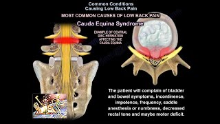
When the patient comes to you for evaluation of low back pain, the first question that you want to ask is if the patient has any bladder or bowel problems. You on a separate low back pain or acute low back pain from cauda equina. Cauda equina syndrome is an orthopedic emergency! It needs to be diagnosed early and treated by surgery early. Make sure that you do not miss cauda equina syndrome. Ask the patient about any bowel bladder or bowel changes. If there is any concern, then you need to get a stat MRI with a wet or stat reading.
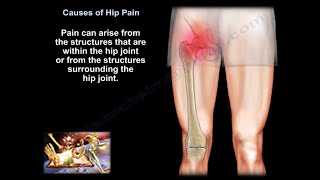
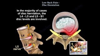
Acute low back pain or low back pain with sciatica radiating to the lower leg and to the foot.
Both conditions are initially treated conservatively for at least 6 weeks by:
Physical therapy
Antiinflammatory medication
Limited activity (as guided by the pain)
Treat the condition conservatively even if there is a big disc herniation on the MRI–wait at least 6 weeks.
Acute Low Back Pain
90% of patients will have symptoms resolve in 1 month.
Smoking, depression, and vibrations will increase the incidence of low back pain
Intradiscal pressure (IDP) changes with different body positions. The lowest pressure is measured while the patient is lying supine. The highest IDP is measured while the patient is sitting, leaning forward and holding weight. If the patient is experiencing low back pain and there is history of cancer, you need to get an xray and MRI, especially if the pain occurs during rest and at night. In case of a renal tumor, you will probably need to do arteriography and embolization of the spinal lesion. The spine is a common location for metastatic tumors. Metastases occur in the vertebral body and goes to the pedicle. Loss of about 30% to 40% of the bone marrow should occur before we can detect the lesion on xray. Loss of the pedicle bone will give us a "weak sign.”
Infection will occur within the intervertebral disc space. The erythrocyte sedimentation rate (ESR) and Creactive protein (CRP) levels will be elevated. Only 50% of the cases will have fever and less than 50% will have an elevated white blood cell count (WBC). Get a blood culture (positive in about 24% of cases). Get an MRI and give antibiotics as guided by biopsy, culture and sensitivity. If the patient has an epidural abscess, you will do surgery, especially if there is a deterioration of the neurological function. If there is an infection postoperatively, you can diagnose it with a high Creactive protein (CRP).
Osteoporotic spine fracture
Osteoporotic bone is at risk of fracture at the wrist, of the spine, and then the hip. If you have an osteoporotic spine, you need to treat it before leads to a hip fracture later on. One fracture of the spine will lead to more spine fractures. After 1 year of treatment with medication, the incidence of vertebral fracture is decreased by 60%. After 2 years incidence of vertebral fracture is decreased by 40%.
When you are dealing with a patient with low back pain:
Treat the patient conservatively.
Do not get an xray in the first 4 to 6 weeks unless there are some "red flags "such as:
Patient is older
Patient has a metastatic tumor/history of cancer
Infection is suspected
Patient has trauma
Osteoporotic fracture due to steroid use
You may see an xray that looks like ankylosing spondylitis. Check the SI joint because ankylosing spondylitis starts in the SI joint. You may get HLA–B27. You will find that there are marginal syndesmophytes with diffuse ossification of the disc space without large osteophyte formation. Ankylosing spondylitis is different from Diffuse Idiopathic Skeletal Hyperostosis (DISH) which occurs in diabetics. Get an HbA1c test. Sydesmophytes are none marginal and have larger osteophytes. There is diffuse idiopathic skeletal hyperostosis (DISH), which will have flowing ossification along the anterior lateral aspect of the least for continuous vertebra. DISH is not ankylosing spondylitis. You are going to get an MRI of the spine at a certain point, however you need to start first by getting xrays.
MRI may be a problem.
There are abnormal MRIs in asymptomatic patients (there are false positives)
35% patients less than 40 years of age
90% positive MRIs in asymptomatic patients over 60 years of age
MRI with gadolinium dye
Gadolinium will differentiate a disc from a scar
Both granulation tissue and the recurrent disc could lookalike on routine MRI
If there is a vascular enhancement then it is a granulation tissue and you will need to sit tight and not do surgery
If there is no enhancement, then it is a recurrent disc and it is avascular, consider surgry if indicated
I